SKU:EA1-1000270
Complete breathing system in 7 parts
Complete breathing system in 7 parts
ATTENTION! This item ships separately. The delivery time may vary.
Couldn't load pickup availability
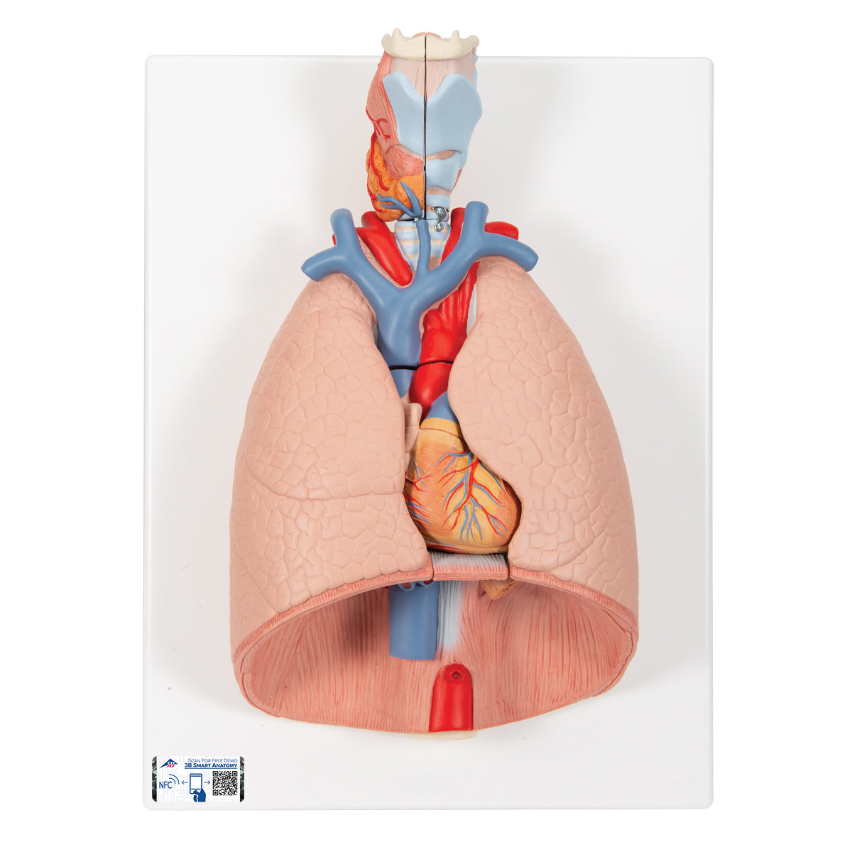
Are you looking for a model that shows all the structures necessary for breathing? With this model, it becomes easy to both understand and explain the relationships between the individual parts of the breathing system and how their interaction allows us to breathe.
The model has the dimensions 41 x 31 x 12 cm (height x width x length), weighs 1.845 kg and is delivered on a white stand. The model is mounted on the stand so that the lungs are oriented horizontally, corresponding to a person lying down.
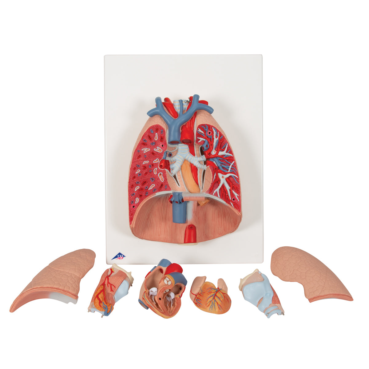
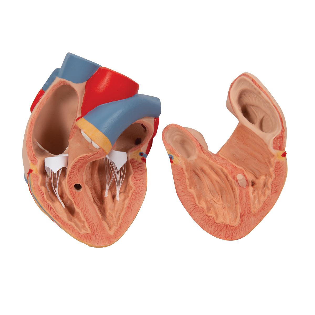
The model can be divided into 7 parts, as the heart, throat and front part of each lung can be removed. The heart can be further divided so that the inside of the heart chambers can be seen.
Anatomically speaking
Anatomically speaking
Anatomically, the model shows the entire respiratory system starting from the throat (oral cavity, nasal cavity and pharynx are not included). The following items are displayed:
Throat
The trachea and bronchial tree
The heart
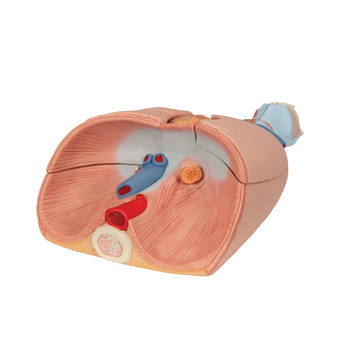
Diaphragm
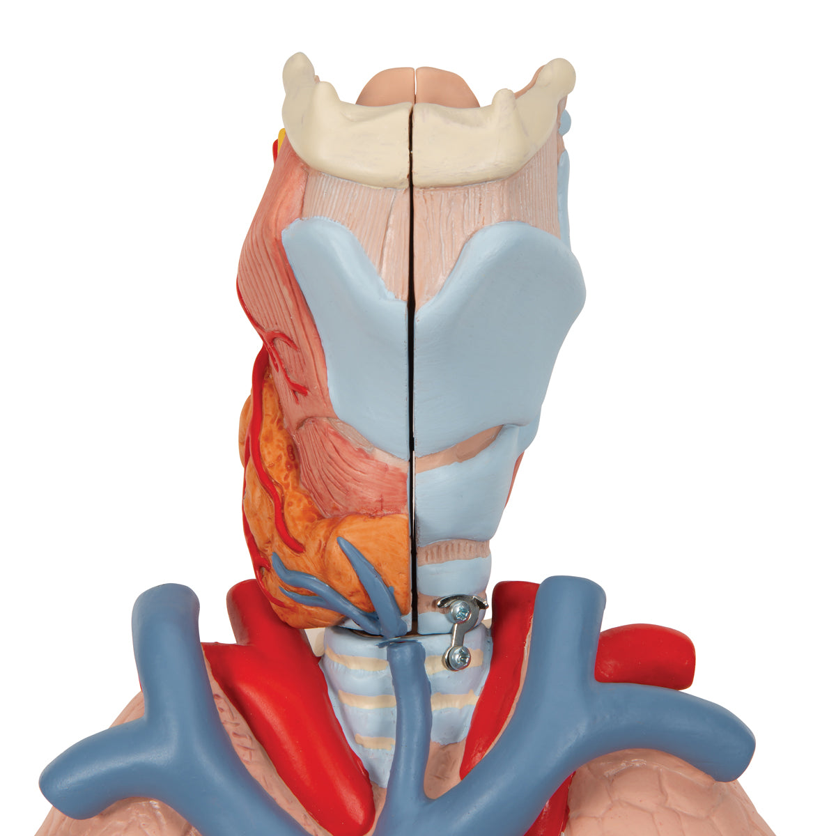
The throat (larynx) is shown with its various cartilages and membranes, as well as their relationships to the bone of the tongue (os hyoideum) and the thyroid gland (glandula thyroidea).
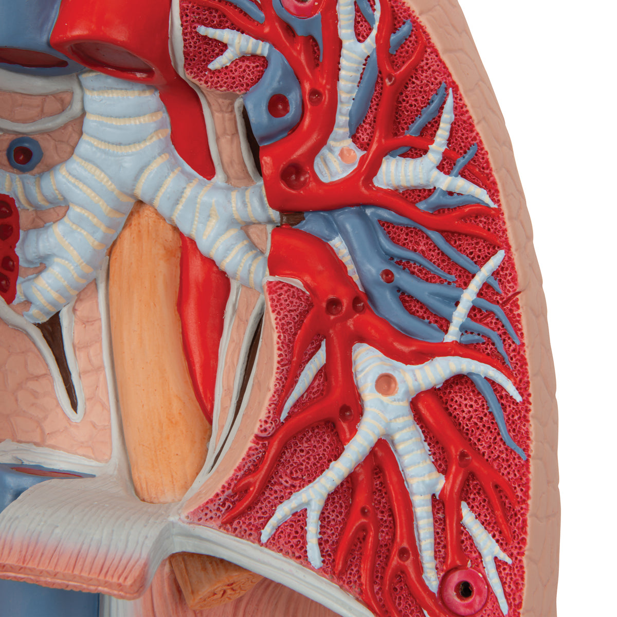
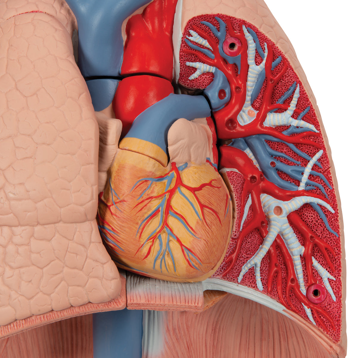
The model shows the division of the trachea into main bronchi, lobe bronchi and segmental bronchi. Likewise, it is seen how A. pulmonalis and V. pulmonalis follow the bronchial tree into the lung tissue.
The heart is imaged with all the vessels that run respectively to and from the heart and the coronary arteries. All four chambers of the heart are visible, as well as the valves located between the atria, ventricles and outflow vessels (respectively A. pulmonalis and aorta).
In addition, it is seen how the lungs and heart rest on the diaphragm, and how the oesophagus, aorta and inferior vena cava pass through each of their openings in the diaphragm on their way down into the abdominal cavity.
Flexibility
Flexibility
Clinically speaking
Clinically speaking
Clinically speaking, the model can be used to understand diseases in the lungs. Examples are blood clots in the lungs, pneumothorax, obstructive lung diseases, bronchiectasis and cancer of the lungs.
The model can also be used to understand disease elsewhere in the chest cavity and on the neck. Examples of heart disease are valvular disease, inflammation and blood clots.
Furthermore, the model can be used to understand examination methods such as bronchoscopy or the installation of a tracheostomy.
Share a link to this product

A safe transaction
For 19 years I have been managing eAnatomi and sold anatomical models and posters to 'almost everyone' who has anything to do with anatomi in Scandinavia and abroad. When you place your order with eAnatomi, you place your order with me and I personally guarantee a safe transaction.
Christian Birksø
Owner and founder of eAnatomi and Anatomic Aesthetics

