SKU:EA1-1000294
Skin model of 3 skin areas. Can be separated into 3 parts
Skin model of 3 skin areas. Can be separated into 3 parts
Out of stock
this product is made to order. To place an order please call or write us.Couldn't load pickup availability
This skin model shows the skin of the human scalp, palm and back of the hand.
The model weighs 1.8 kg and measures 34 x 39 x 15.5 cm. It shows the skin at 80 times normal size and can be separated into 3 parts, which are held together by magnets. It is delivered on a stand (white plastic sheet).
Anatomical features
Anatomical features
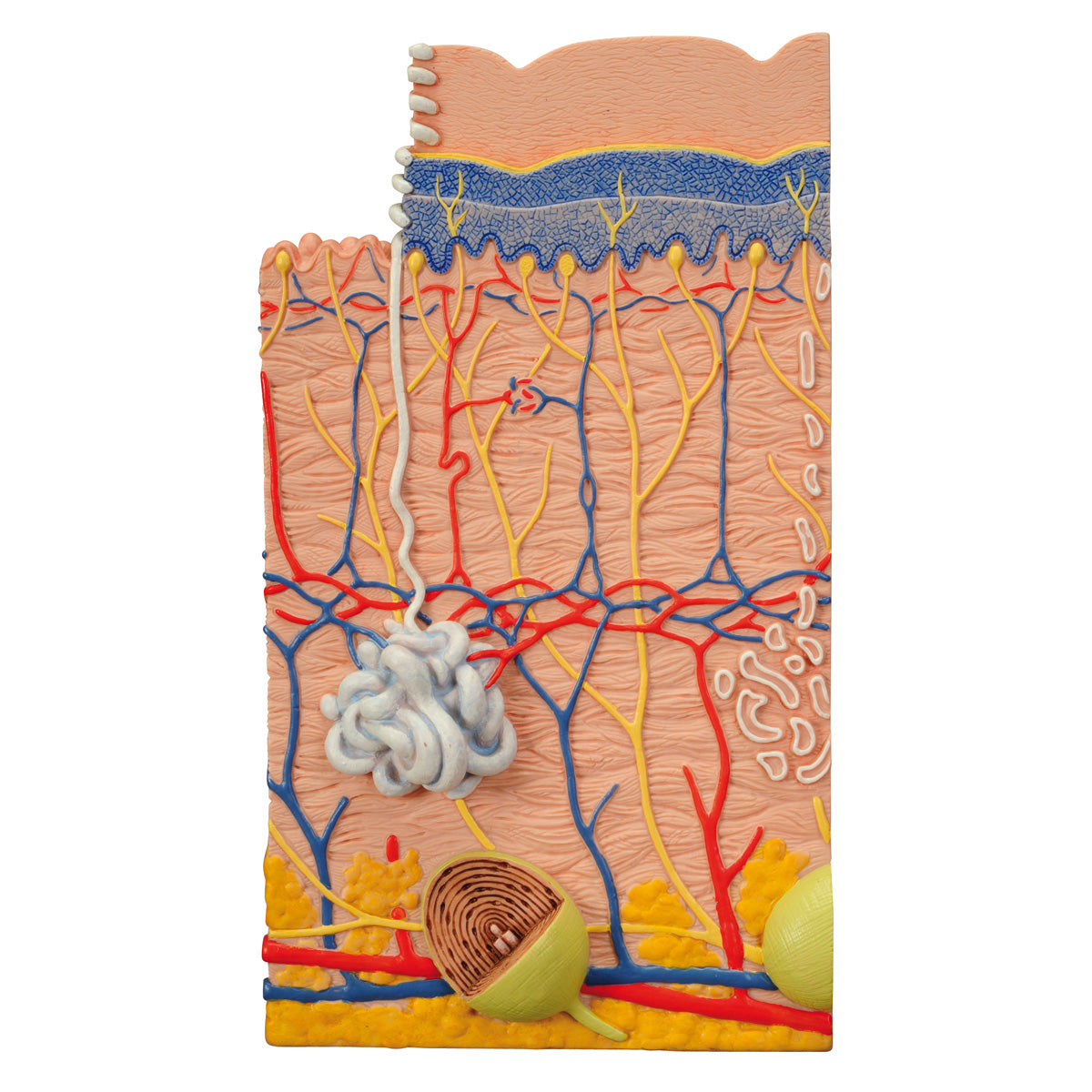
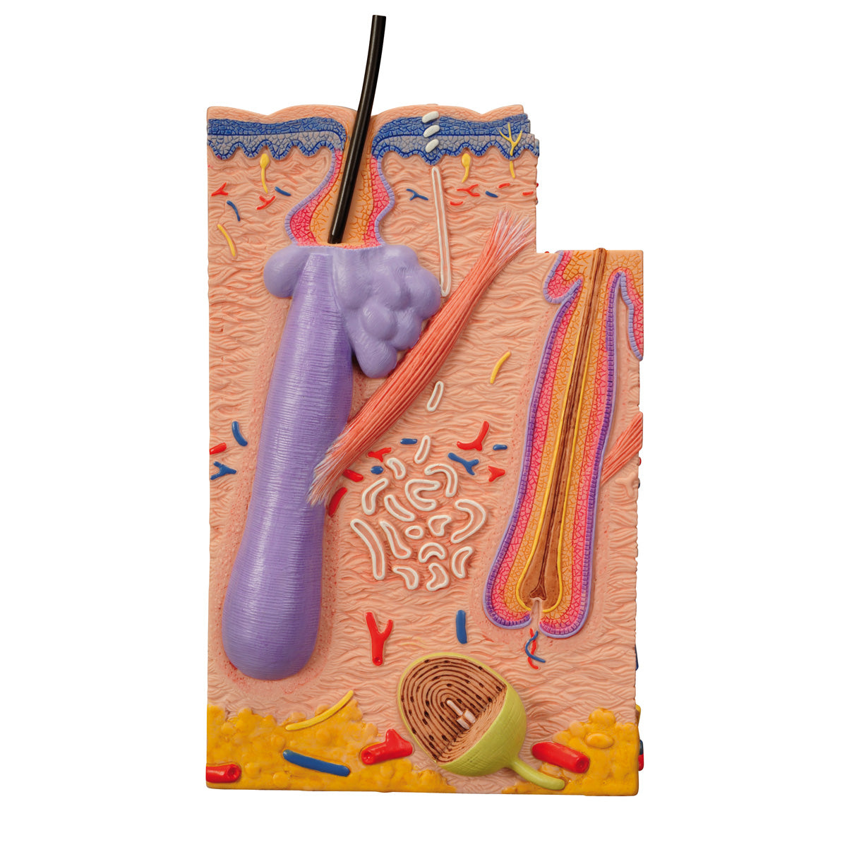
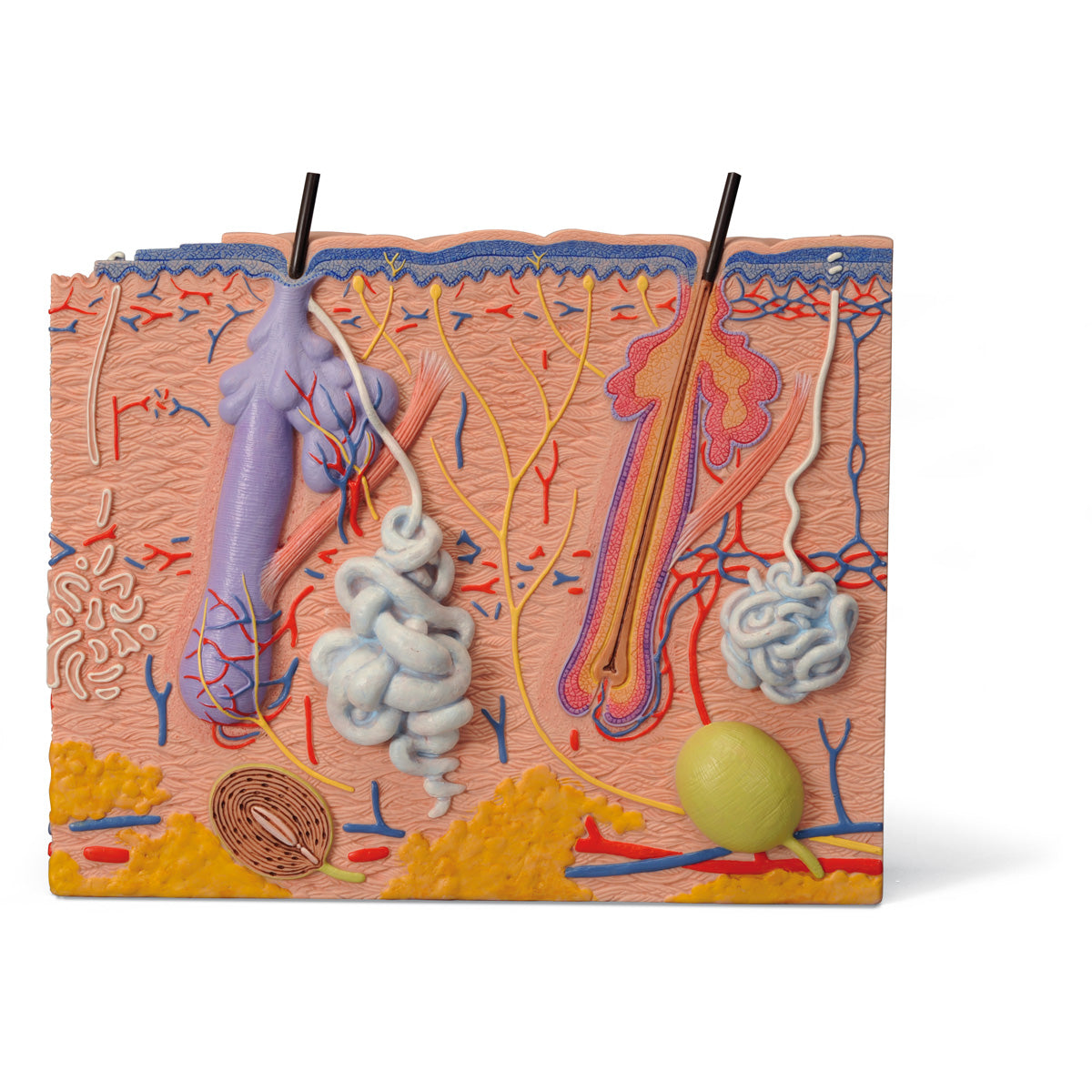
Anatomically, the model shows hair and the skin's two layers, the epidermis and dermis. The skin (subcutis) is also seen.
The epidermis consists primarily of keratinocytes, which create a layered structure due to their displacement upwards towards the skin surface. On this skin model, much of the epidermis is shown in blue, but the individual layers (such as the stratum granulosum and stratum spinosum) are difficult to identify. The basement membrane can be seen at the bottom.
In the dermis, the thick connective tissue layer with nerves, blood vessels, hair follicles with associated smooth muscle (m. arrector pili) and sweat and sebaceous glands can be seen.
Some of the subcutaneous tissue is also shown in yellow at the bottom, which symbolizes fat cells (since the subcutaneous tissue mainly consists of fat cells).
The model can be separated into 3 parts, so that the skin from 3 different parts of the body can be studied. The different parts can be seen from different angles (including from above). You see the following:
A part where you can see hairless skin and extra thick epidermis ("thick skin"), which characterizes the skin in e.g. the palm of the hand. Seen from above, friction ridges/papillary ridges can also be seen
A part where you can see skin with hair. Seen from above, you can also see polygonal or diamond-shaped fields in the epidermis, which characterizes the skin of e.g. the back of the hand
A part where you can see many anatomical details in the hair root and the hair follicle, which characterizes the skin in e.g. the scalp
Product flexibility
Product flexibility
Clinical features
Clinical features
Clinically, the model can be used to understand skin disorders and skin diseases such as psoriasis, folliculitis, acne and skin tumors.
It can also be used to understand other things, such as burns, which are traditionally divided into degrees (1st degree, 2nd degree, etc.).
Share a link to this product

A safe deal
For 19 years I have been at the head of eAnatomi and sold anatomical models and posters to 'almost' everyone who has anything to do with anatomy in Denmark and abroad. When you shop at eAnatomi, you shop with me and I personally guarantee a safe deal.
Christian Birksø
Owner and founder of eAnatomi ApS


