


SKU:EA1-A276
Flexible pelvic model showing the pelvic floor muscles and related tissues in the female
Flexible pelvic model showing the pelvic floor muscles and related tissues in the female
Out of stock
this product is made to order. To place an order please call or write us.Couldn't load pickup availability
If you are looking for an educational pelvic model where the focus is on the muscles etc. in the woman's pelvic floor, this model is ideal. It comes with mobility in the SI joints (sacroiliac joints) as well as numbering and associated naming.
The pelvic model is particularly popular with customers who want an explanatory model in connection with rehabilitation after childbirth, operative interventions, etc.
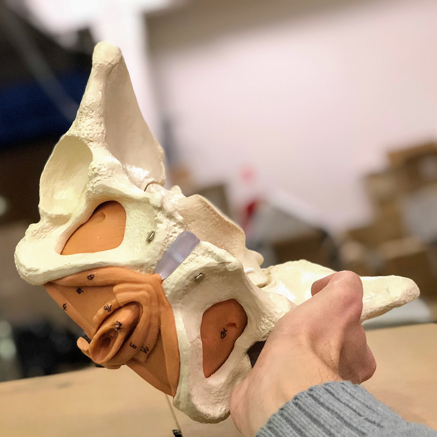
It is developed in natural adult size, and the level of detail on the bones is good. The model cannot be separated, but both the SI joints and the joint between the sacrum and tailbone are slightly movable because they are fitted with elastics. The front joint (symphysis), on the other hand, is not movable at all. Below you can read more about the pelvic measurements in relation to the pelvis as a way of birth.
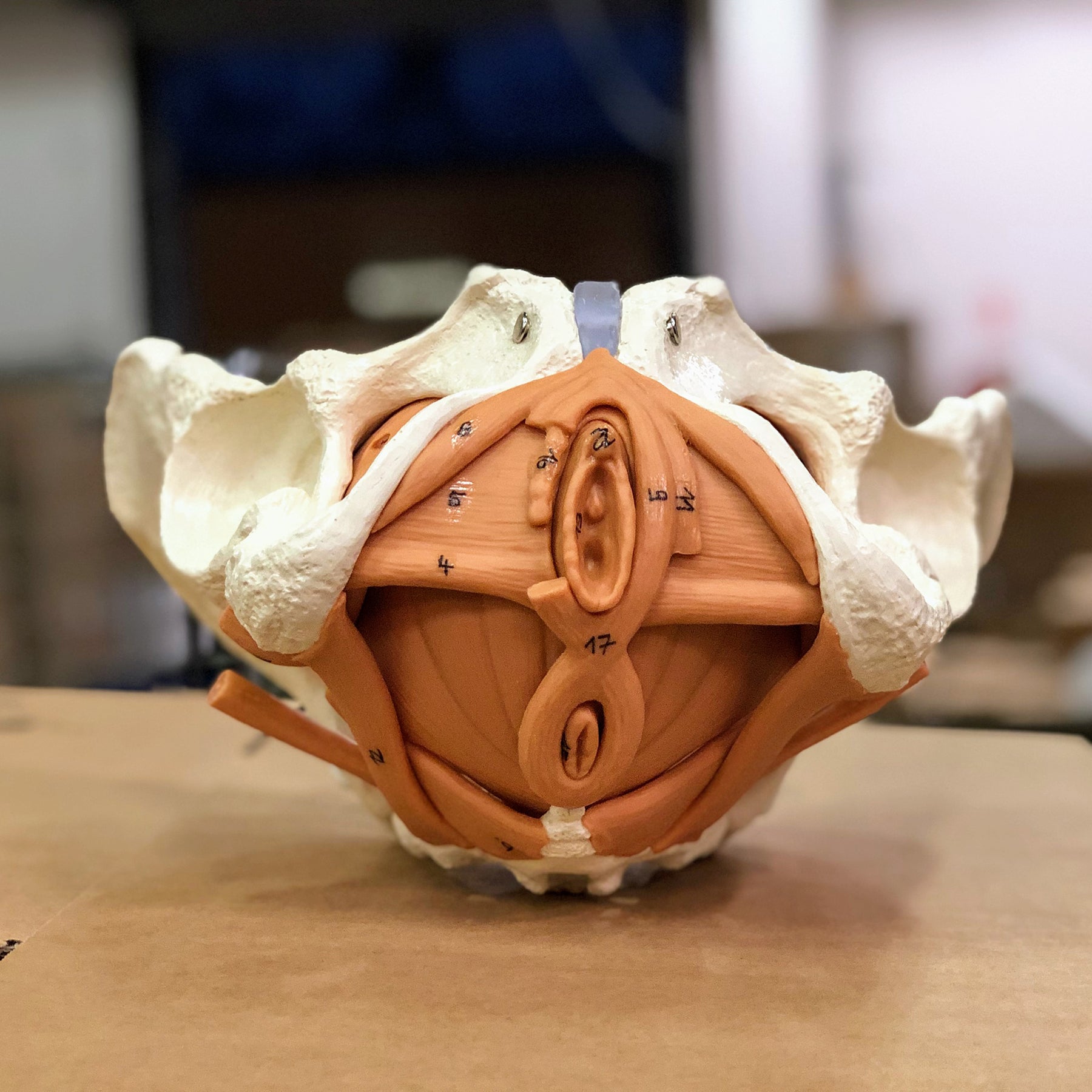
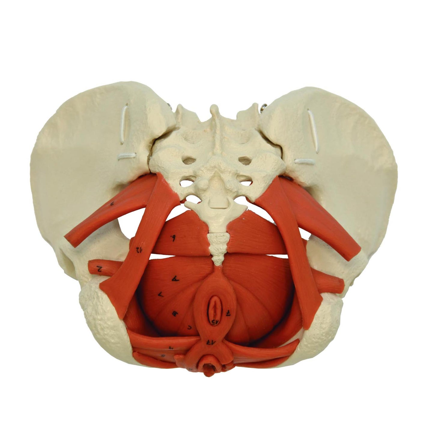
NB: It must be emphasized that the color of the muscles is brown, as can be seen in the pictures from our warehouse on the left. In the manufacturer's product images, the muscles unfortunately appear in orange (e.g. in the first image on the left).
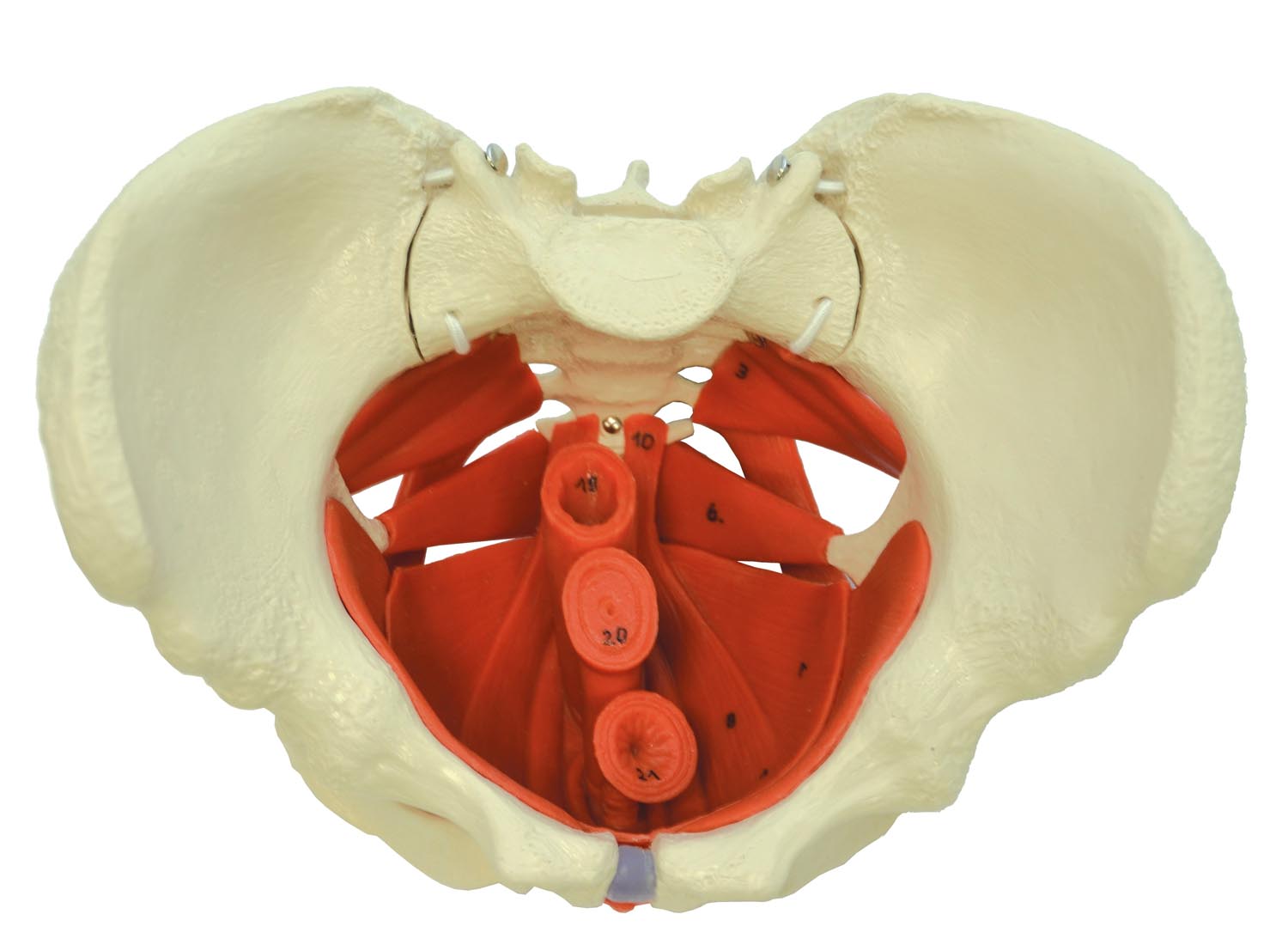
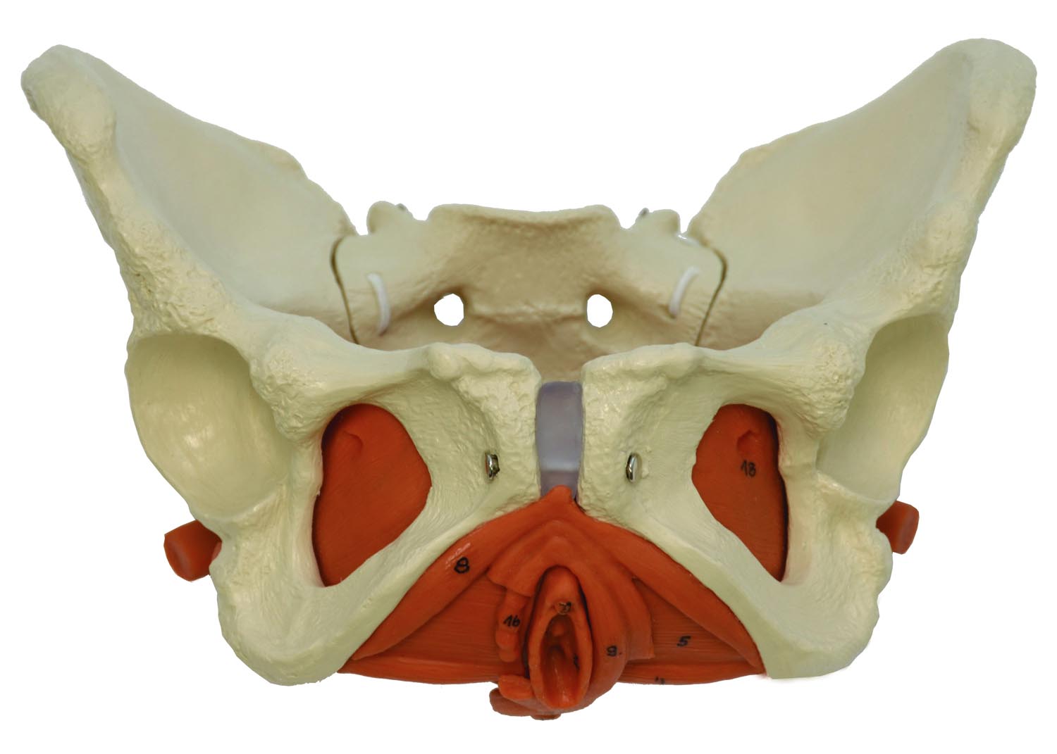
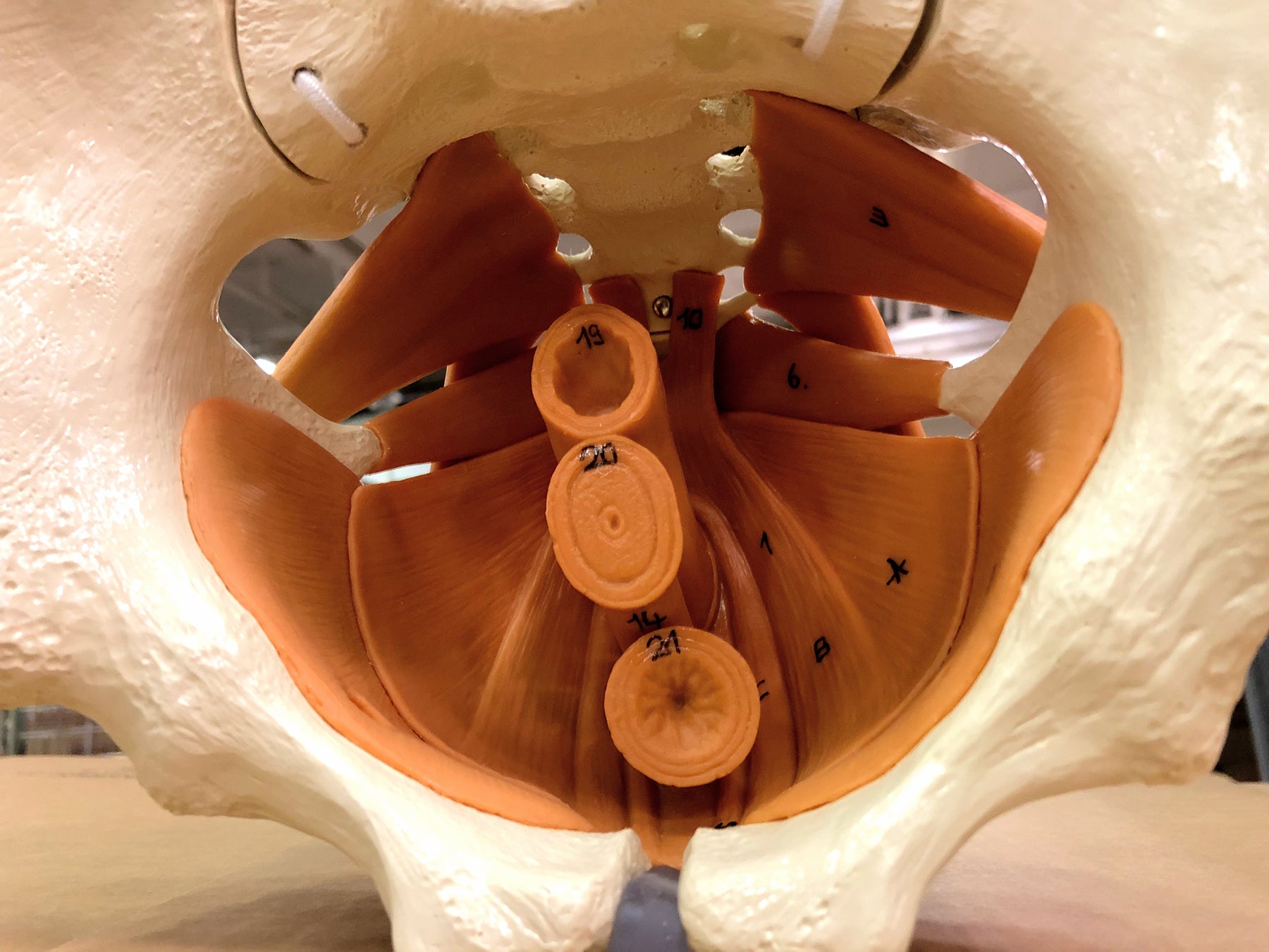
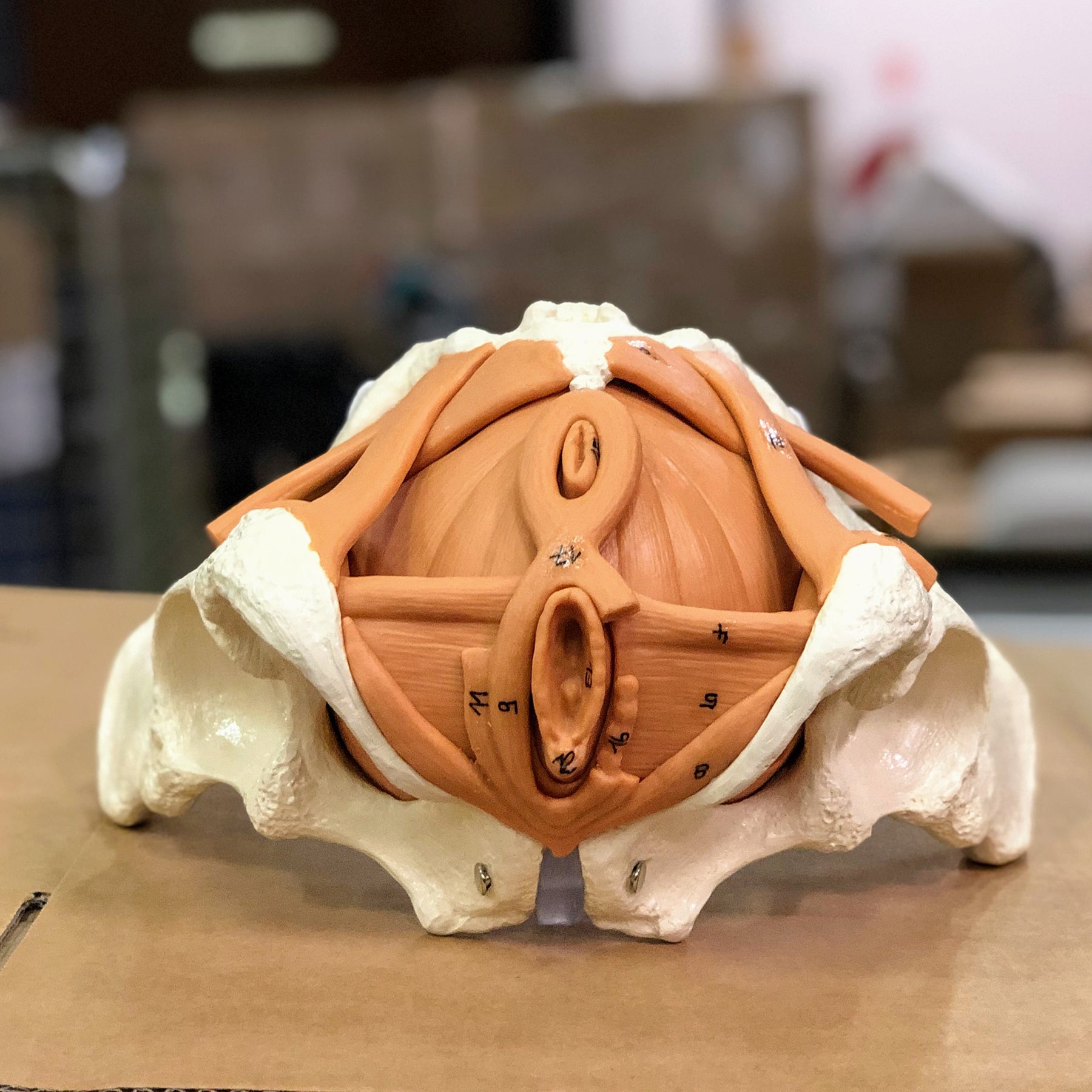
Important anatomical structures are numbered on the model (muscles, etc.). The purchase does not include a detailed overview with naming cf. the mentioned numberings, but you can instead download and print the overview via the link on the left under the product images (click on the PDF symbol). The nomenclature is drawn up in Latin.
Anatomical features
Anatomical features
Anatomically, the model shows the woman's pelvis and the muscles in the pelvic floor. Also seen a bit of the genitals, a bit of the urinary bladder with the urethra and a bit of the rectum.
The pelvis consists of the 3 building blocks, which are the 2 hip bones and the sacrum. Overall, the model shows the following bones:
Os coxae (the 2 hip bones) of which both are composed
- Os ilium (iliac bone/hip bone)
- Os ischii (seat bone)
- Os pubis (pubic bone)
Us sacrum (sacrum) incl. os coccygis (coccyx)
In the bone tissue, the most important details can be seen, such as large nodules. Smaller details such as the linea glutaea are omitted. Furthermore, a single ligament/ligament (like sacrotuberale) is seen, which has the same color as the muscles.
The pelvic floor (also called the pelvic floor or diaphragma pelvis in Latin) consists of the following muscles, which are clearly visible:
- M. levator ani (consisting of m. pubococcygeus and m. iliococcygeus)
- M. coccygeus
Other muscles are also seen such as the ischiocavernosus muscle, the bulbospongiosus muscle, the obturator internus muscle and the piriformis muscle.
The internal and external genitalia are only partially visible. They are seen in relation to the urinary bladder (vesica urinaria) with the urethra (urethra) and the rectum (rectum). Apart from the urethra, these organs are also only partially seen.
The pelvis as a birth canal
Normally the following 3 connection lines are specified:
- Conjugata vera (normally measures 11 cm in the female pelvis)
- Diameter obliqua (usually measures 12.5 cm in the woman's pelvis)
- Diameter transversa (usually measures 13 cm in the woman's pelvis)
On this pelvis model, the above measurements are respectively: 10.5 cm, 13 cm and 14.5 cm.
You can also use a measure for the anteroposterior exit from the pelvis, which is the distance from the symphysis to the tip of the coccyx. On this model, the distance is approximately 11 cm (but it depends on the position of the coccyx, as it is movable).
Product flexibility
Product flexibility
In terms of movement, the model's joints must be mentioned. Both the SI joints (articulatio sacroiliaca) and the joint between the sacrum and coccyx (articulatio sacrococcygea) are flexible because they are held in place by elastics. Little movement can therefore be demonstrated in these joints.
The front joint called the symphysis (symphysis pubica), on the other hand, is not movable at all.
It must be emphasized that the model is not designed to be disassembled in any way.
Clinical features
Clinical features
Clinically speaking, the model as mentioned above can be used in connection with rehabilitation after birth, operative interventions etc. It can also be used in connection with birth preparation and the like.
Furthermore, it can be used to understand pelvic fractures with malposition as well as specific diseases such as axial spondylarthritis, which i.a. involving the SI joint.
Share a link to this product


A safe deal
For 19 years I have been at the head of eAnatomi and sold anatomical models and posters to 'almost' everyone who has anything to do with anatomy in Denmark and abroad. When you shop at eAnatomi, you shop with me and I personally guarantee a safe deal.
Christian Birksø
Owner and founder of eAnatomi ApS