Skip to product information
1 of 6

SKU:EA-1000144

Flexible model of the cervical spine with the brainstem, spinal nerves and a. vertebralis

Flexible model of the cervical spine with the brainstem, spinal nerves and a. vertebralis

Regular price $119.00 USD
Regular price Sale price $119.00 USD
Sale Sold out
Shipping calculated at checkout.

Out of stock

this product is made to order. To place an order please call or write us.

This flexible model shows the cervical vertebrae, brainstem, spinal nerves, a. vertebralis and the neck bone (os occipitale).


The model measures 19 cm in height and weighs approximately 0.3 kg. All the cervical vertebrae are held together by a copper wire, and in addition, an elastic system has been inserted into the upper cervical vertebrae. The model is delivered on a white stand.

Anatomical features

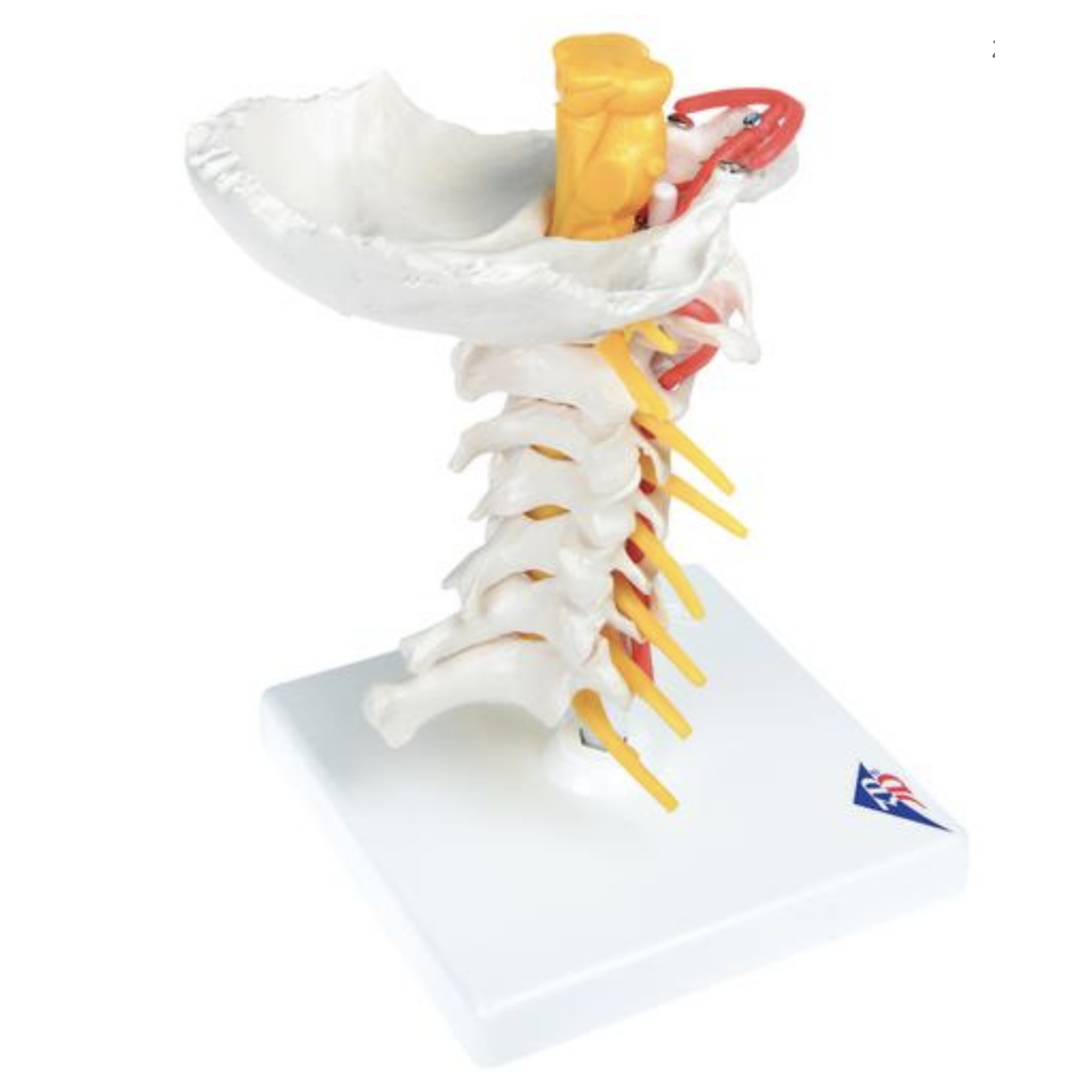
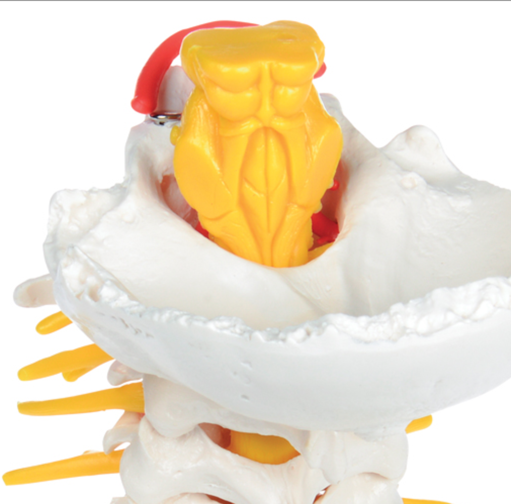
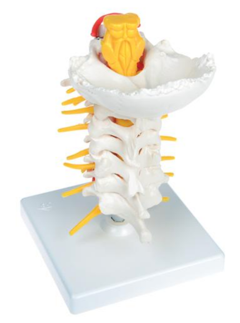
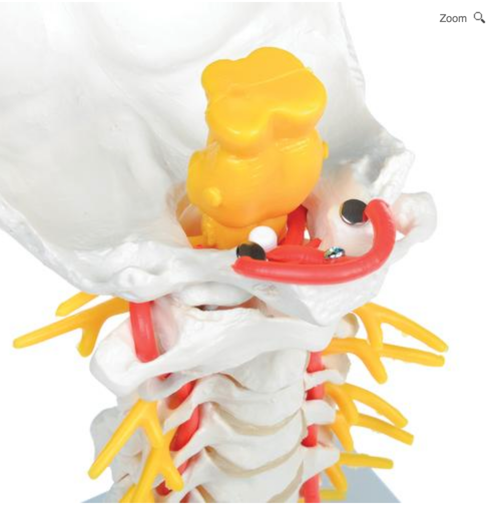
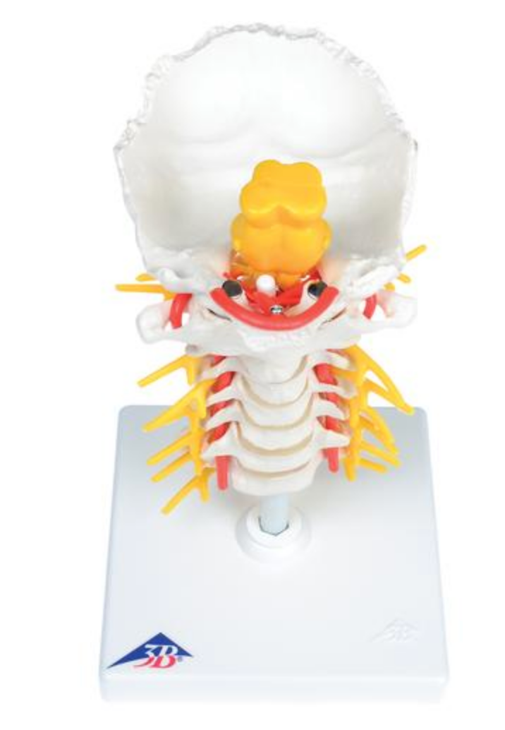
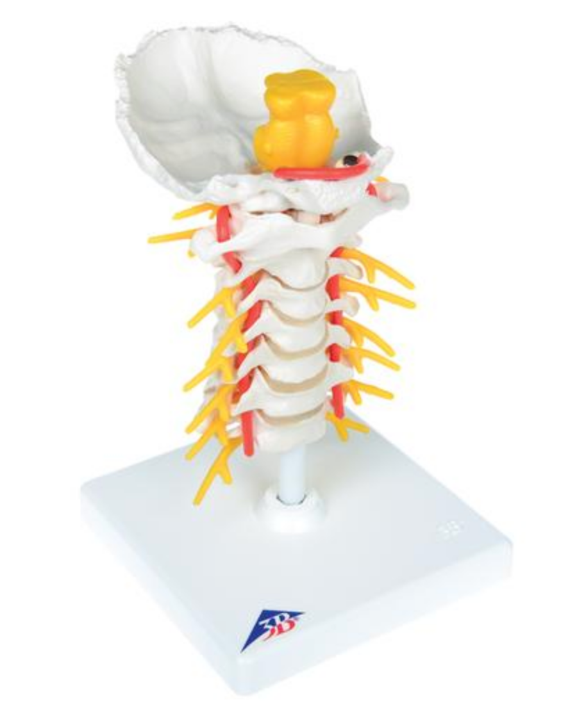
Anatomically, the model shows all 7 cervical vertebrae as well as the neck bone (os occipitale). Therefore, the model shows the two most important joint types in the spinal column, which are the symphyses (disci/band disks) between the vertebral bodies and the facet joints, which are sliding joints between the vertebrae's pivots. Furthermore, both upper and lower neck joints are visible.


The model also shows the brainstem (with the midbrain, pons and medulla oblongata), spinal nerves (with both the anterior and posterior radix) and the vertebral artery. Overall, the bone quality is good, which is why, for example, larger protrusions are seen in the bone tissue.

Product flexibility

In terms of movement, the model is flexible. The mobility of the human spine is great in the cervical region (which is lordotic). This model can be used to demonstrate the most important movements, which are flexion-extension, lateral flexion and rotation.


The neck bone (os occipitale), upper cervical vertebra (atlas) and second upper cervical vertebra (axis) are gathered together with elastic string, which causes a special elastic effect, where the vertebrae both follow each other during movement and come together again in a natural way. You can thus demonstrate the most important movements in the neck joints, which are the nodding and rocking of the head, which takes place in the upper neck joint and the shaking of the head, which takes place in the lower neck joint.

Clinical features

Clinically, this model is ideal for understanding disc herniation, root affection and spondylolisthesis (slippage) of the cervical spine. It can also be used to understand disorders such as spondylosis, spinal stenosis, osteoarthritis, vertebral artery dissection and fractures.

View full details

A safe deal

For 19 years I have been at the head of eAnatomi and sold anatomical models and posters to 'almost' everyone who has anything to do with anatomy in Denmark and abroad. When you shop at eAnatomi, you shop with me and I personally guarantee a safe deal.

Christian Birksø
Owner and founder of eAnatomi ApS

Read more about eAnatomi