SKU:EA1-1019311
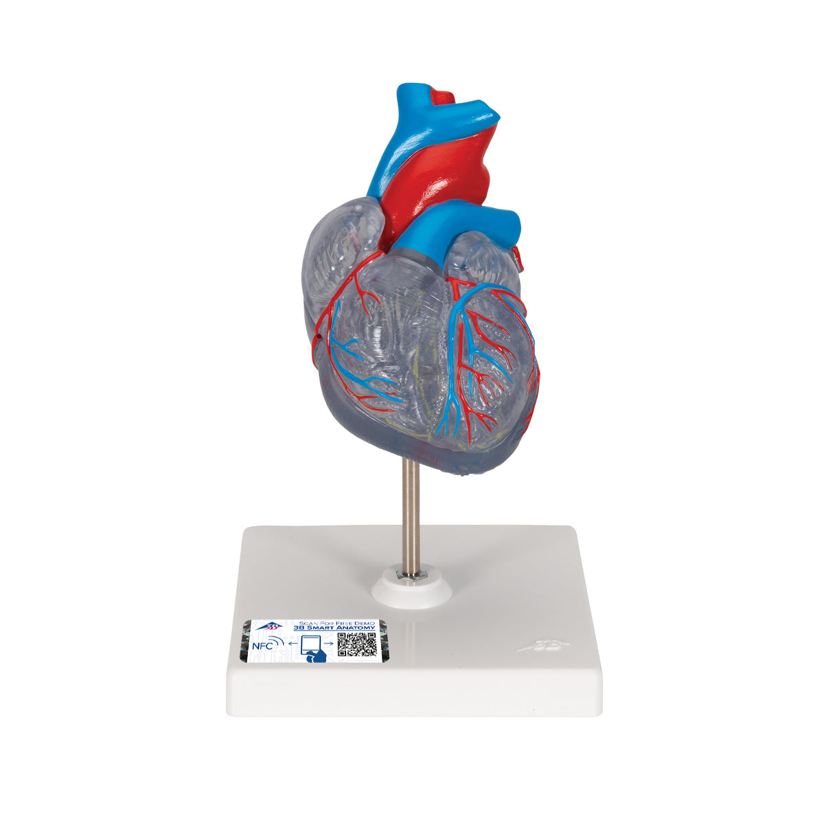
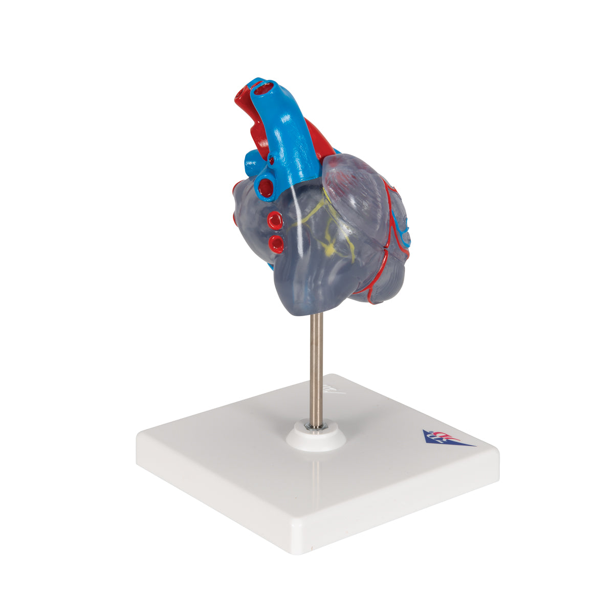
Scaled and transparent heart model with the impulse conduction system
Scaled and transparent heart model with the impulse conduction system
Out of stock
this product is made to order. To place an order please call or write us.Couldn't load pickup availability
Anatomical features
Anatomical features
Anatomically speaking, the following are emphasized on the model:
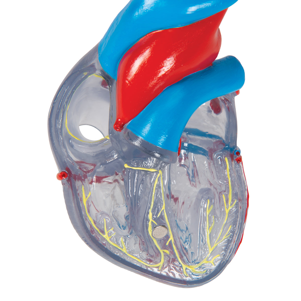
1) The impulse line system
2) Coronary arteries and coronary veins
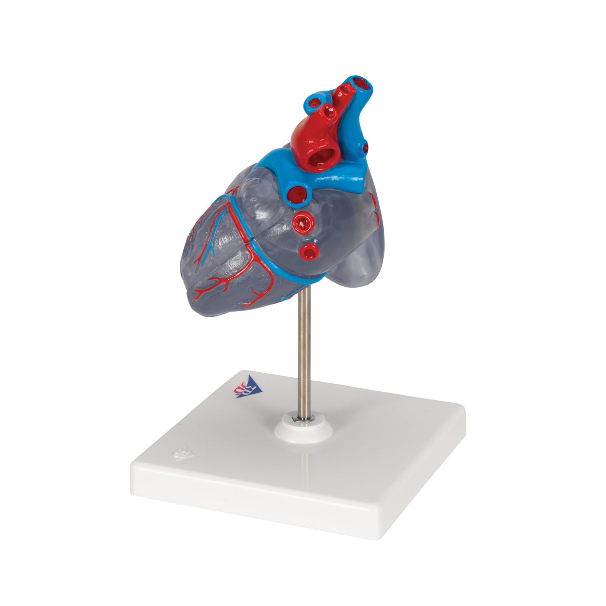
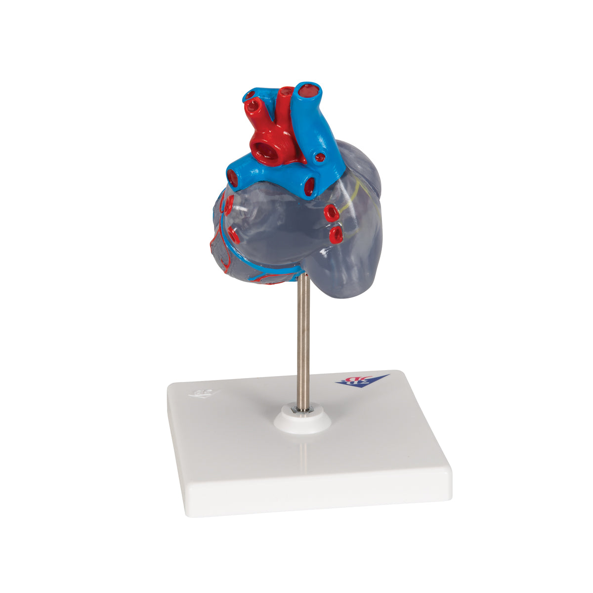
3) The large vessels that carry blood to and from the heart
Although it also illustrates the chambers of the heart, they appear less clear. Smaller anatomical structures are not included.
The impulse conduction system is shown in an extremely educational way, so that you can see the process from the sinus node onwards through the other important structures such as the Atrioventricular node, bundle of His, crura and Purkinje fibers.
The model primarily shows the large coronary arteries and the coronary vein. Arteries are illustrated in red and veins in blue.
The right coronary artery, called arteria coronaria dexter (or RCA) runs right around the heart to the back surface and gives off the ramus interventricularis posterior, which is also seen. The left heart artery is called arteria coronaryia sinister. Its course to the left is also seen. It divides into the two large vessels called ramus interventricularis anterior (or LAD) and ramus circumflexus (or CX).
Most of the venous blood collects in the coronary vein (sinus coronarius), which empties into the right atrium. Smaller veins are also seen.
The large vessels that carry blood to and from the heart include the aorta (body artery), v. cava superior (upper vena cava), truncus pulmonalis (pulmonary artery/pulmonary artery). You can also see the openings to the v. cava inferior (lower vena cava) and vv. pulmonales (pulmonary veins).
Product flexibility
Product flexibility
Clinical features
Clinical features
Clinically speaking, the model does not show anything pathological (disease-related) but is ideal for elucidating conduction disturbances such as atrial fibrillation (atrial fibrillation), AV block and bundle branch block. It is also ideal for the illumination of atherosclerosis and blood clots in the coronary arteries as well as the medical treatment or invasive cardiology such as balloon dilation (PCI) and CAG.
Share a link to this product

A safe deal
For 19 years I have been at the head of eAnatomi and sold anatomical models and posters to 'almost' everyone who has anything to do with anatomy in Denmark and abroad. When you shop at eAnatomi, you shop with me and I personally guarantee a safe deal.
Christian Birksø
Owner and founder of eAnatomi ApS

