SKU:EA1-1000222
Anatomical brain model in 2 parts
Anatomical brain model in 2 parts
Out of stock
this product is made to order. To place an order please call or write us.Couldn't load pickup availability
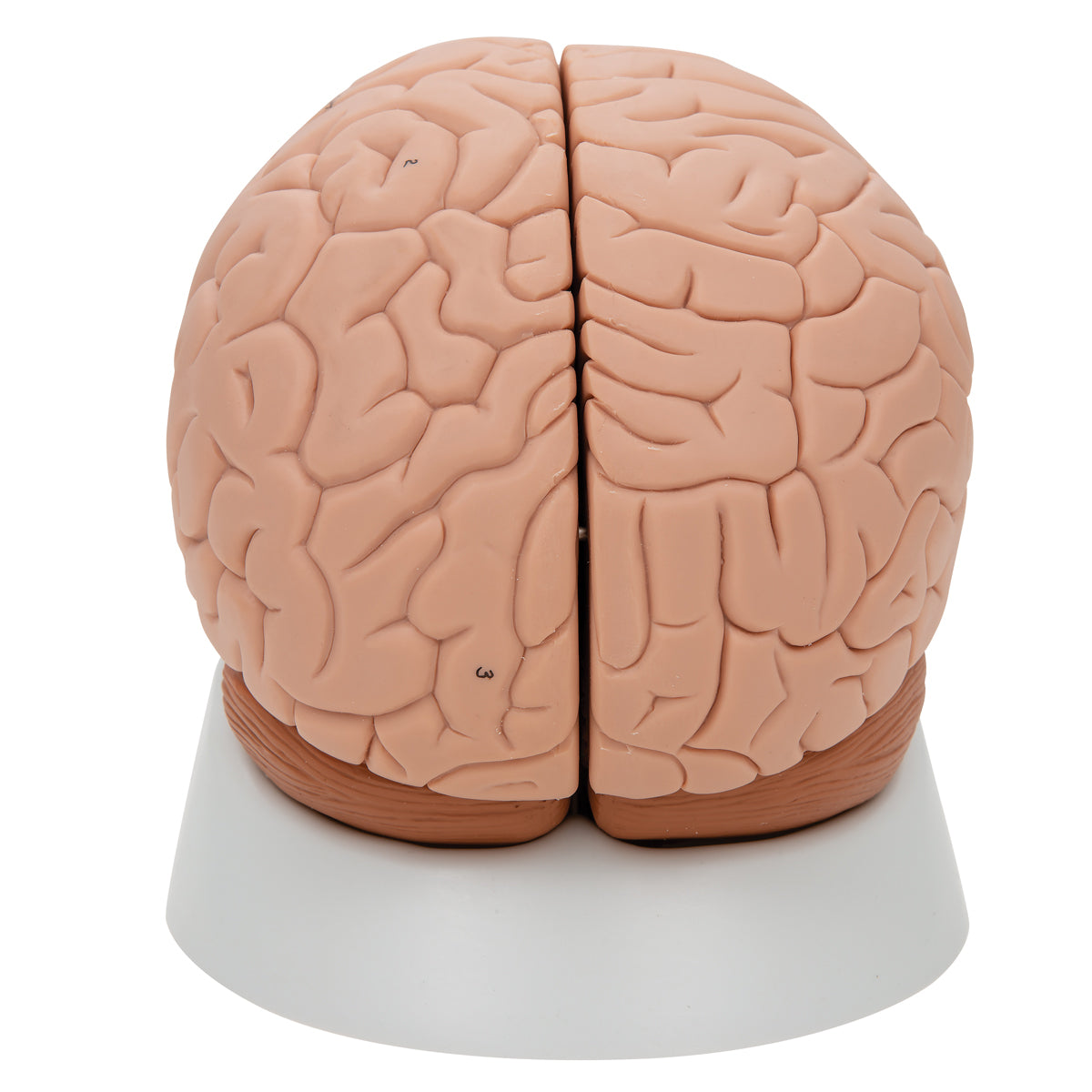
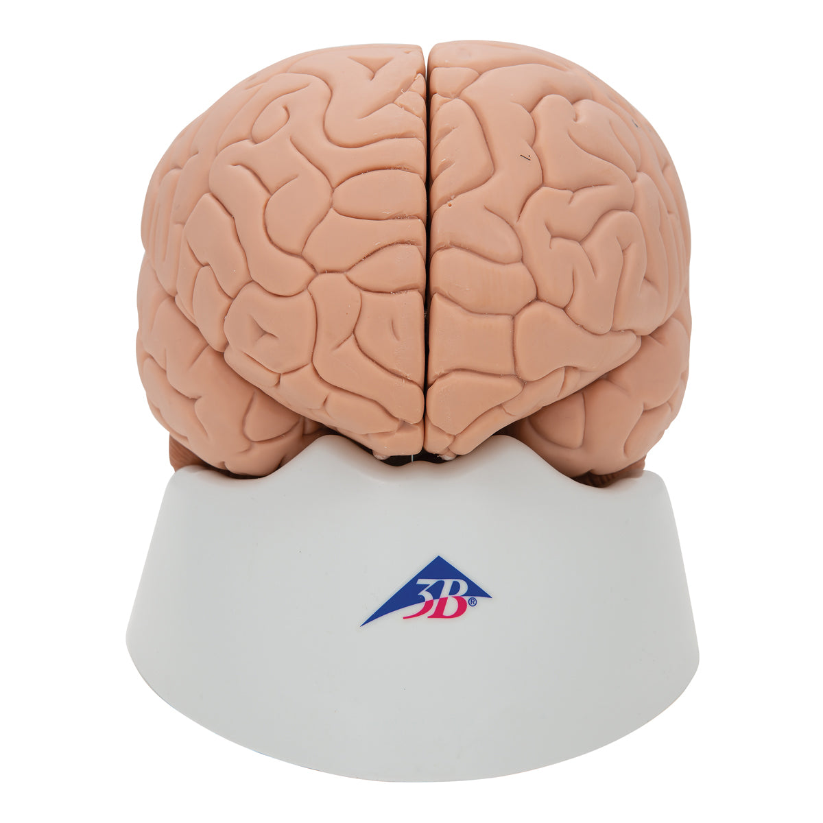
If you are looking for a simple brain model that can simply be split in half and does not look too lifelike, this model is ideal.
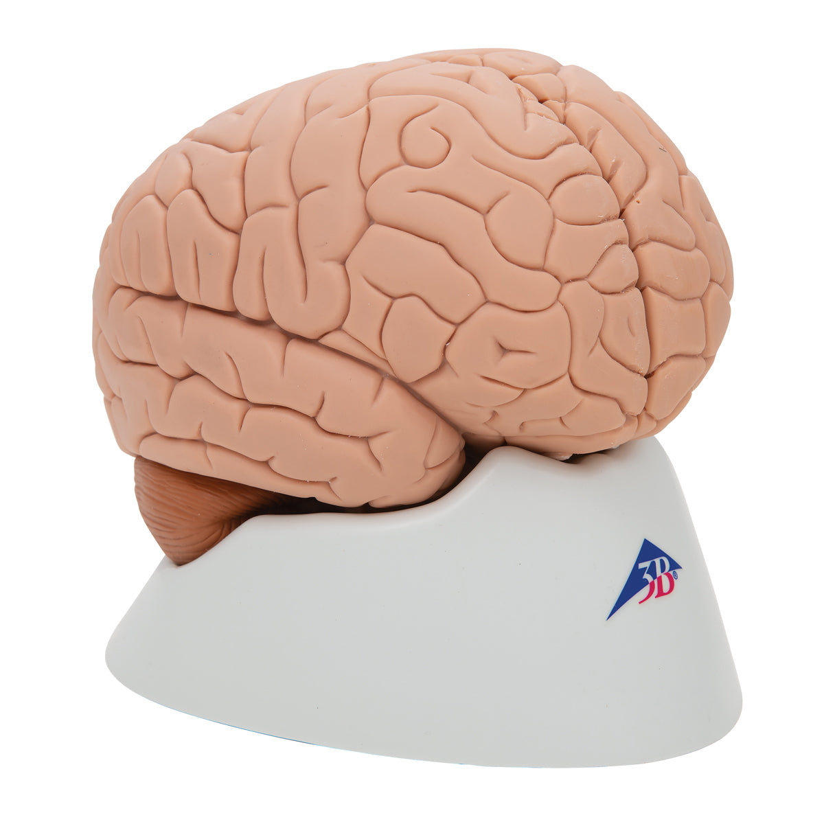
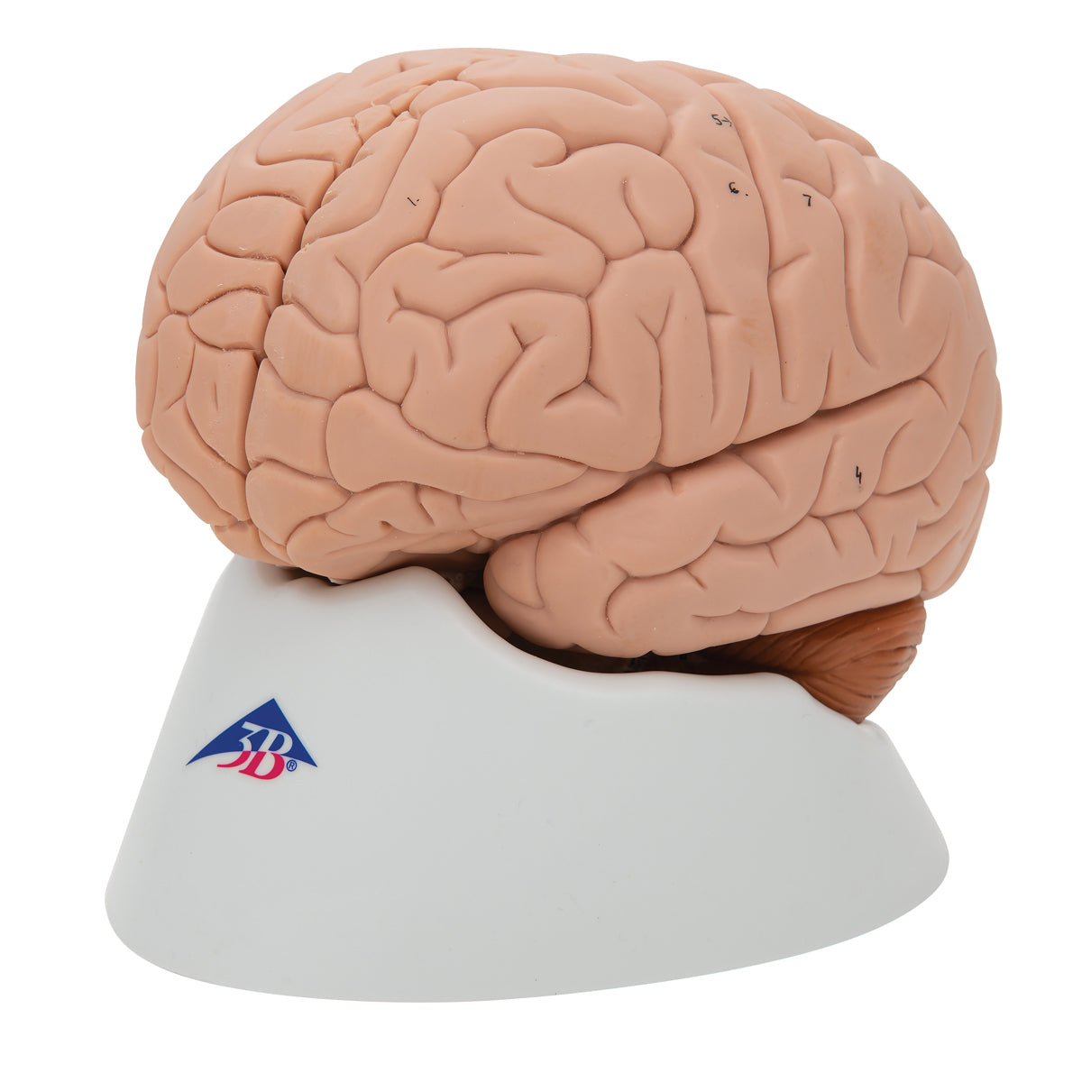
The brain model is cast in hollow and solid plastic, which makes the material flexible because it can both be squeezed quite a bit and moved a bit. This makes the model pleasant to touch and work with when it needs to be taken apart and studied.
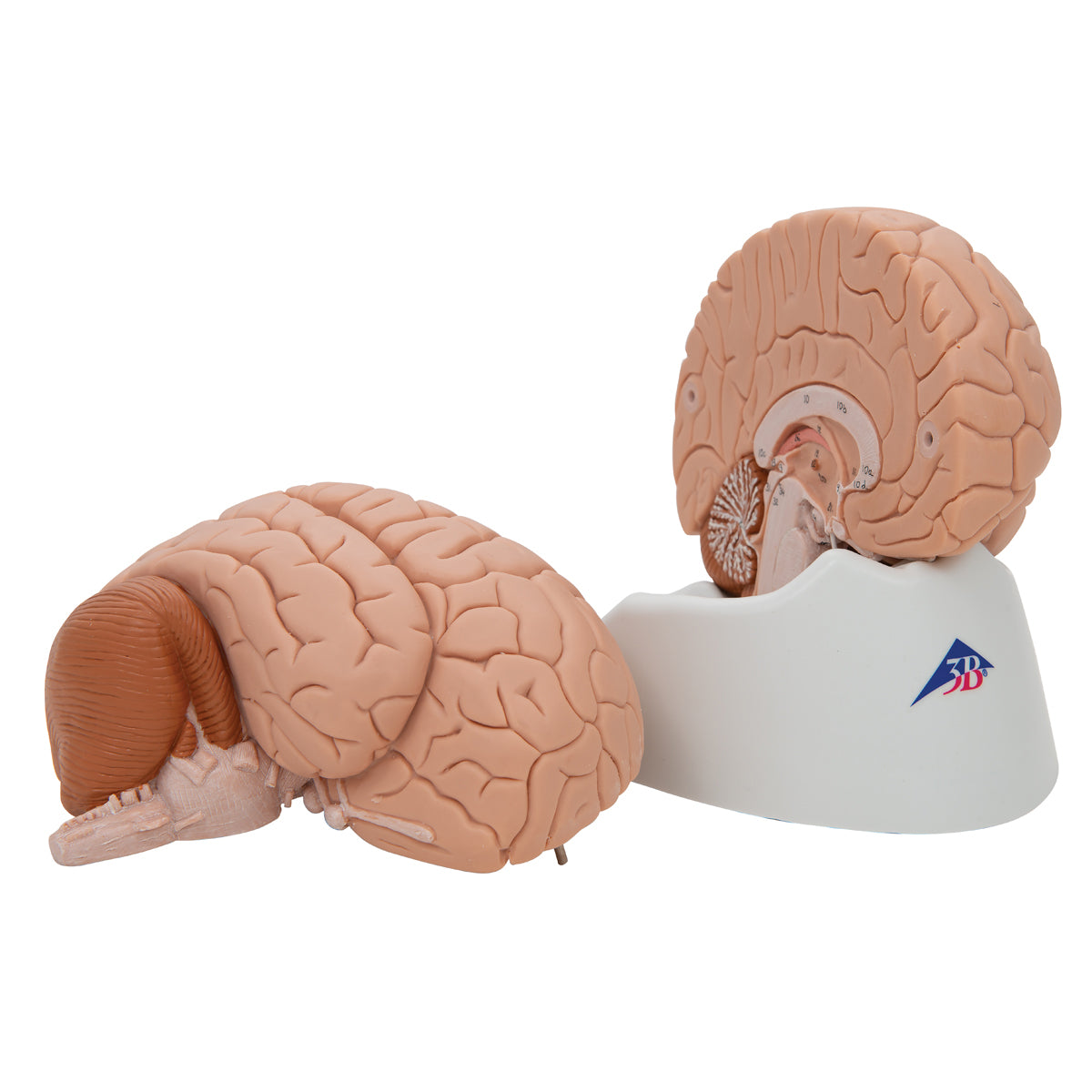
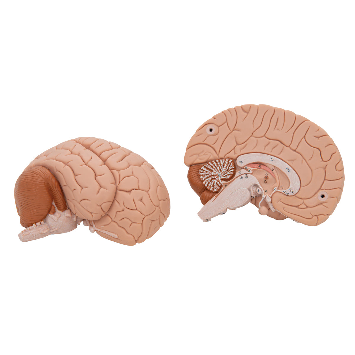
Different areas of the brain appear in different shades. Below you can read more about the anatomical details such as the limbic system. It can be split in the middle, and the 2 parts are held together via small metal pins. The size of the model corresponds to the brain of an adult person. The dimensions are 15 x 14 x 17.5 cm. The weight is approximately 800 grams and it comes on a white removable stand.
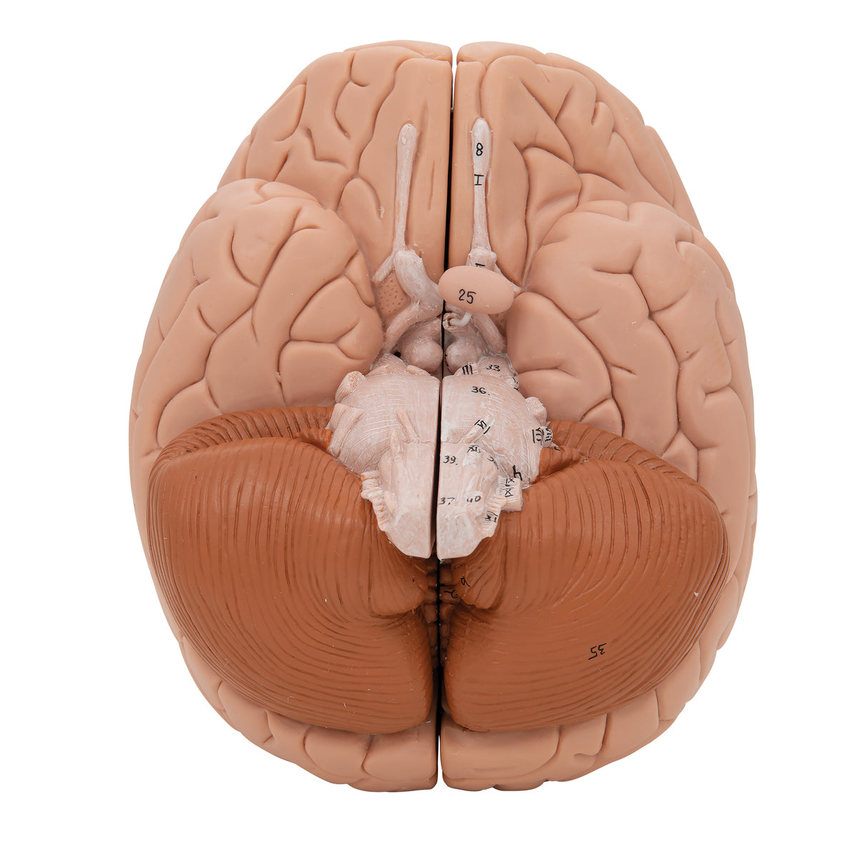
Important anatomical structures are numbered on the model.
NB: The numbering and naming are indicative. Therefore, be critical in your use, as figures for an anatomical structure may, for example, be located on the border of another structure.
Anatomical features
Anatomical features
Anatomically, the model shows the human brain, which can generally be divided into the cerebrum (cerebrum), the cerebellum (cerebellum) and the brain stem (truncus encephali).
These 3 structures are clearly separated via different color tones, but the difference between gray and white matter cannot be seen on this model (however seen in the cerebellum).
In the cerebrum (telencephalon and diencephalon), the lobes of the brain, as well as the thalamus and hypothalamus (and the pituitary gland) are primarily seen
In the cerebellum, the vermis cerebelli and the cerebellar hemispheres (hemisperium cerebelli) are seen
In the brainstem you can see its 3 parts (the midbrain, the pons and the medulla oblongata) as well as the apparent origin of the cranial nerves (also called the cranial nerves)
Other structures are also seen, such as the brainstem, the fornix, a bit of the ventricular system and the first 2 cranial nerves (the olfactory and optic nerves), which do not originate from the brainstem
The limbic system
Many of our customers ask about the limbic system in connection with the purchase of brain models. Hence this description.
The limbic system includes various anatomical structures in the central nervous system (CNS), and is primarily responsible for emotional functions such as anxiety, aggressiveness, mood, memory and social adaptability. Clinically, it is therefore often related to psychiatric disorders.
The limbic system includes, among other things amygdala, hippocampus, gyrus parahippocampalis, hypothalamus, fornix, corpus mammillare, the prefrontal cerebral cortex and the monoaminergic systems of the brainstem. The list is quite a bit longer - especially because numerous fiber connections connect the limbic structures. Many customers ask in particular about the amygdala and hippocampus (which is why they are mentioned first in this section).
NB: In this brain model, neither the amygdala nor the hippocampus can be seen, but some of the other limbic structures can be seen (e.g. the fornix).
The amygdala is involved in anxiety and emotional coloring of sensory impressions. It lies as an almond-shaped nucleus IN FRONT of the hippocampus in the anterior pole of the temporal lobe (amygdala and hippocampus are therefore separate).
The hippocampus is involved in memory. It lies as an irregular twisted structure in the medial part of the temporal lobe.
If one of these 2 structures is to be seen clearly on a brain model, the brain tissue must be shown in a so-called frontal/coronal section through the temporal lobe. The frontal incision roughly corresponds to the incision direction "from ear to ear".
Because the amygdala is IN FRONT of the hippocampus (roughly speaking further forward "toward the forehead"), both of these structures can only be seen on a brain model if the model includes at least 2 frontal sections through the temporal lobe - or if the brain model is partially transparent.
We have not yet seen a brain model that shows 2 cuts through the temporal lobe, so that both the amygdala and the hippocampus are seen. In our range, on the other hand, we have a partially see-through brain model in the highest price range, which shows both structures.
All brain models in our range can be separated into different parts. All models (both with and without educational colors) that can be separated into 4 or more parts show the hippocampus. On almost all of these models, the hippocampus is also numbered and named on an overview that can be downloaded from the product descriptions of the brain models.
Product flexibility
Product flexibility
Clinical features
Clinical features
Clinically speaking, the model can be used for a general understanding of lesions and disorders in the areas of the brain that are seen on the model.
Share a link to this product

A safe deal
For 19 years I have been at the head of eAnatomi and sold anatomical models and posters to 'almost' everyone who has anything to do with anatomy in Denmark and abroad. When you shop at eAnatomi, you shop with me and I personally guarantee a safe deal.
Christian Birksø
Owner and founder of eAnatomi ApS