Skip to product information
1 of 14

SKU:EA-A206.3

Advanced skeleton model with muscles in the face, neck and neck, ligaments etc

Advanced skeleton model with muscles in the face, neck and neck, ligaments etc

Regular price $1,229.00 USD
Regular price Sale price $1,229.00 USD
Sale Sold out
Shipping calculated at checkout.
Size

Out of stock

this product is made to order. To place an order please call or write us.

Extra robust skeleton with partially movable back, muscles in the face, neck and neck, ligaments etc

This extra robust skeleton model shows the entire skeleton in natural size from head to toe and is the model in the range that includes the most extra features. The skeleton includes the following:


  • Visible muscles in the left part of the face, the entire neck and neck
  • Possibility to study the teeth individually as well as their anchoring in the jawbone
  • Visible ligaments at joints on the right side
  • Illustrations of the origin and attachment of skeletal muscles using red and blue color on the left side
  • Naming of muscles and bones in accompanying document
  • Vertebral artery
  • The spinal nerves
  • A lumbar disc herniation
  • A partially flexible spine
  • Possibility to "open" the sacrum (os sacrum) and see the course of the spinal nerves


Mounted on a stand, the plastic skeleton measures approx. 180 cm in height. The bones are made of a heavy plastic material, which provides particularly good durability and resistance to breakage. Therefore, it is suitable for frequent moving and rough treatment in, for example, a classroom. Furthermore, the bone color is slightly yellowish, which makes the skeleton appear more realistic.


The level of detail on the bones is really good, and virtually all joints are held together by metal buckles/metal, which allows limited ability to demonstrate the natural movements of the joints, as well as the ability to completely dismount (for example, one arm). Joints on the right side are primarily held together by brown rubber ligaments, and movement in almost all of these joints is severely limited. On the other hand, the spine is partially flexible, and the movement is described below.


The skeleton rests on a solid stand, which can be easily moved because at the bottom it consists of a 5-branched column base with a corresponding number of wheels. A simple dust bag is also included.


As mentioned above, the origin and attachment of skeletal muscles are illustrated in red and blue and numbered. The visible muscles of the face, neck and neck are also numbered, but the visible ligaments are not. A comprehensive list with their names in Latin and English is included (a total of 326 numbers: 120 for skeletal muscles and 206 for bones).

Anatomical features

Anatomically, the skeleton shows all the bones of the body (including the tongue bone and sesamoid bones). Cartilage in the chest and teeth in the upper and lower jaw are also included.


The skull can be quickly disassembled and divided as follows:


The skull cap can be removed
The lower jaw can be removed
A small part of the lower jaw bone can be opened via "a flap"
31 teeth can be extracted


If the mandible is opened via the "flap", tooth roots and nerve canals can be studied. A "toppled" wisdom tooth is also seen.


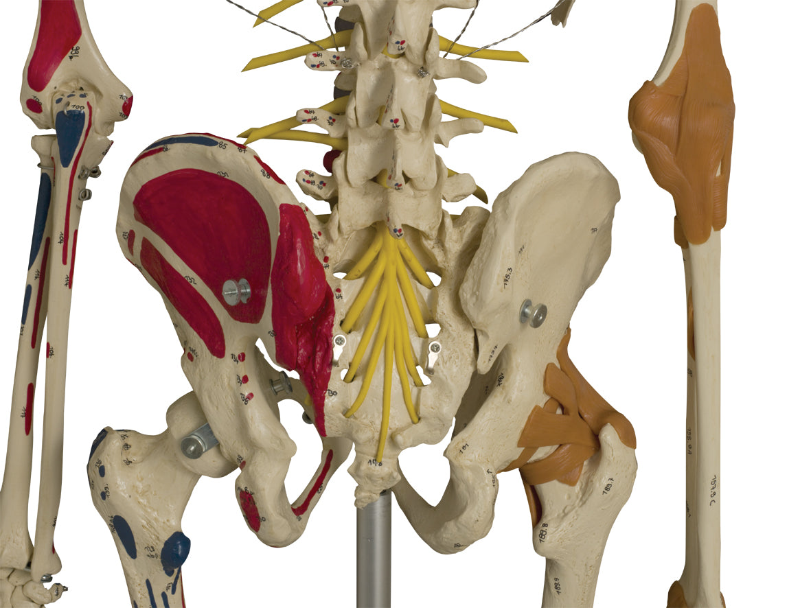
The sacrum (os sacrum) can also be opened and studied. It is thus possible to see the canalis sacralis as well as the Y-shaped canals in its lateral wall (see the images on the left).


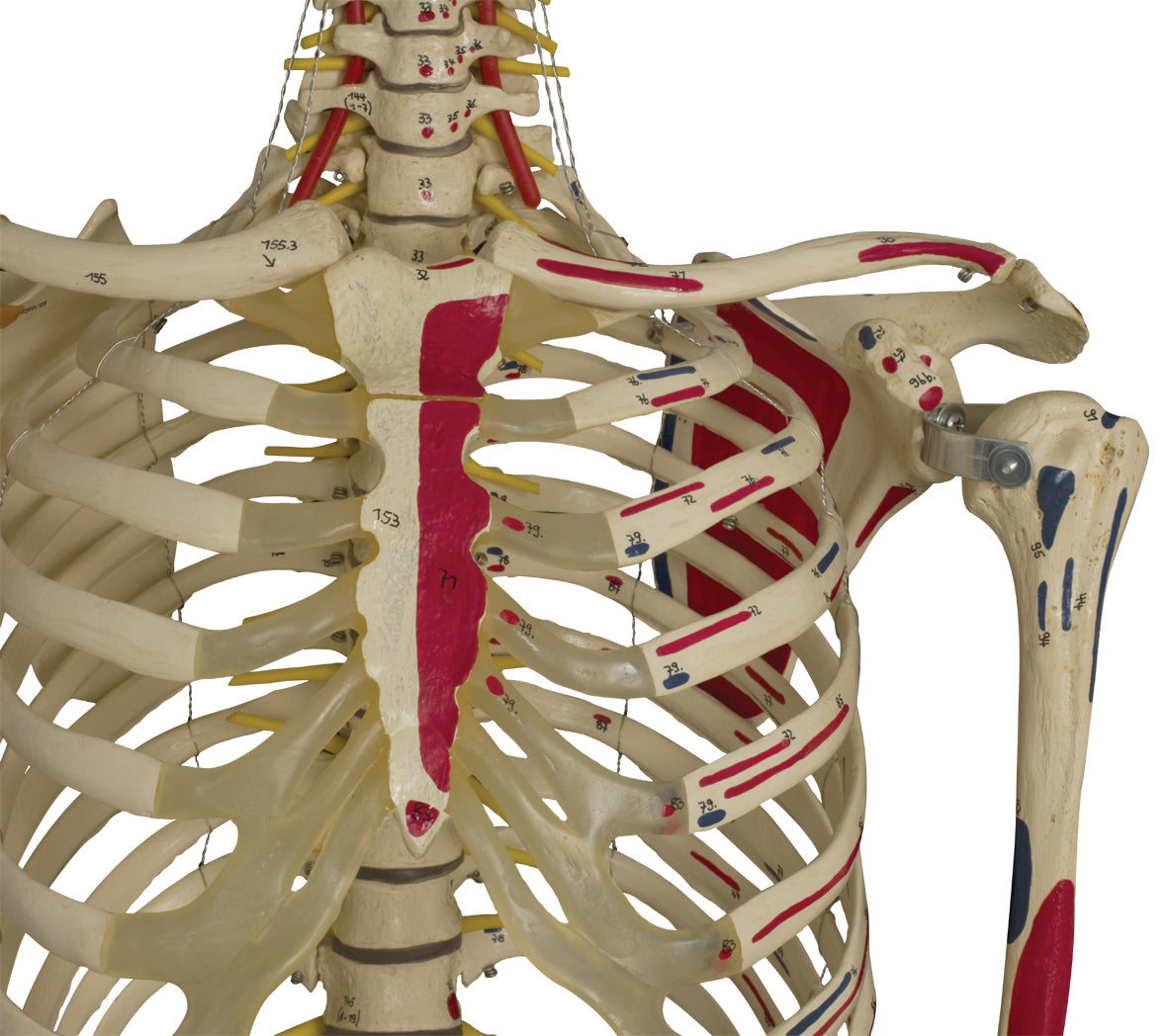
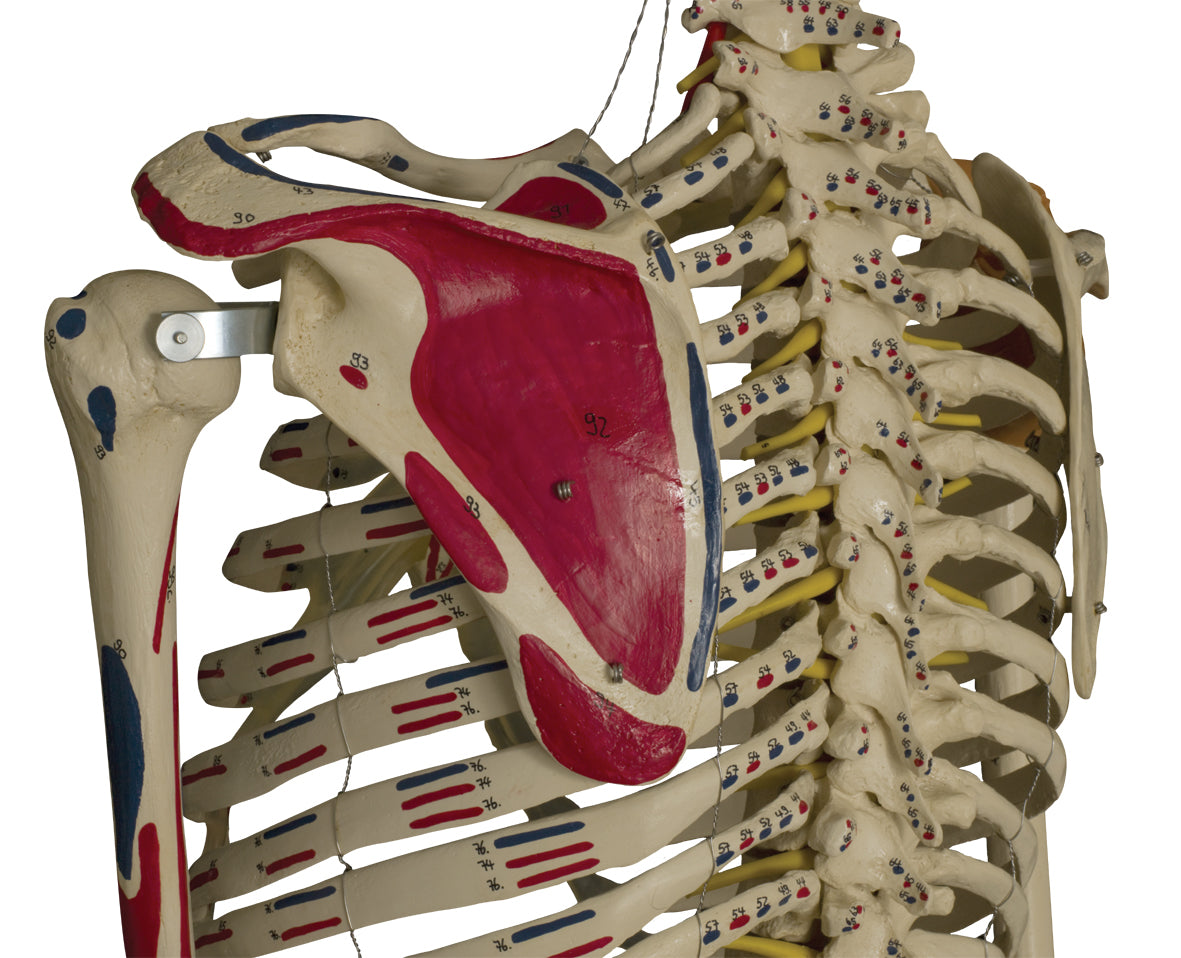
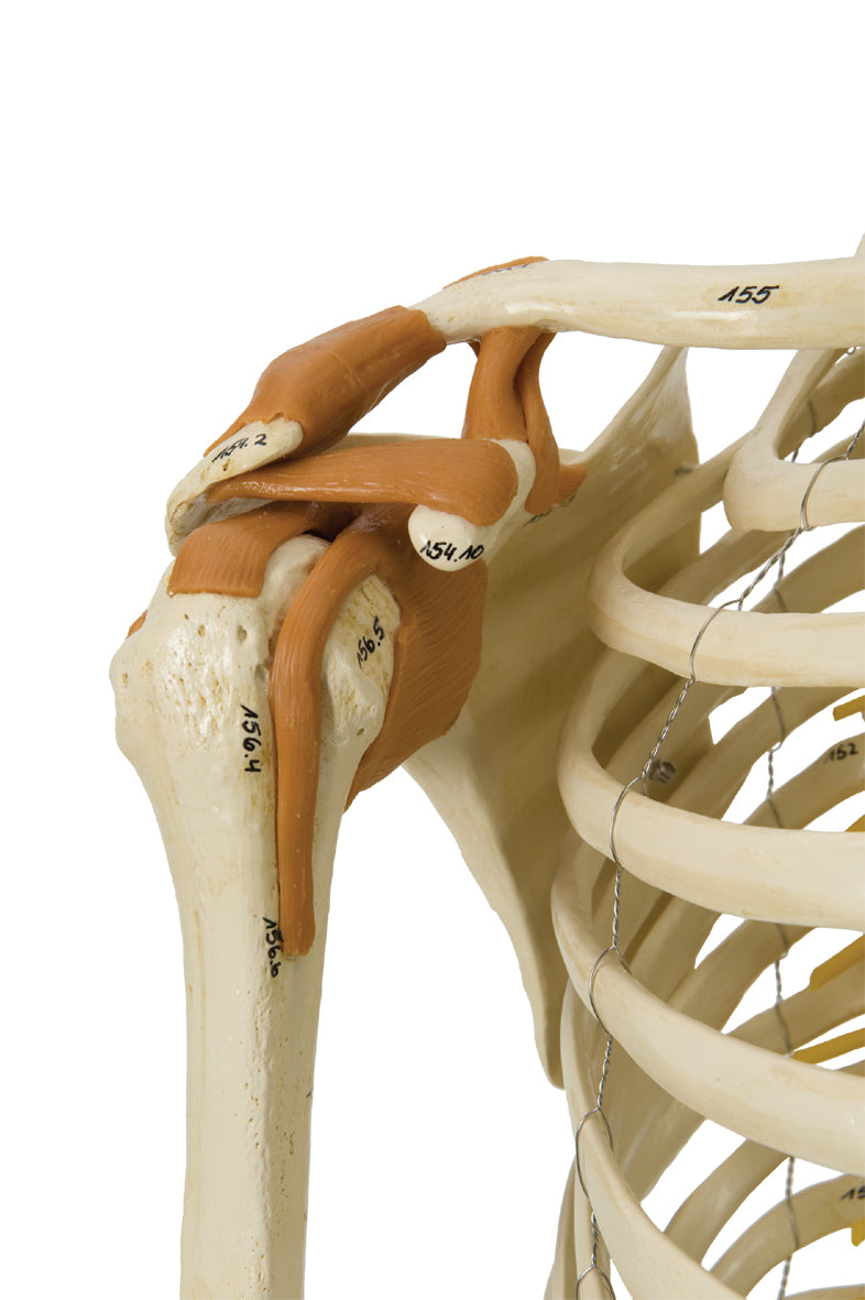
The level of detail on the bones is really good. The scope of "osseous landmarks" includes both large details such as the tuberculum majus and minus on the humerus (upper arm bone).


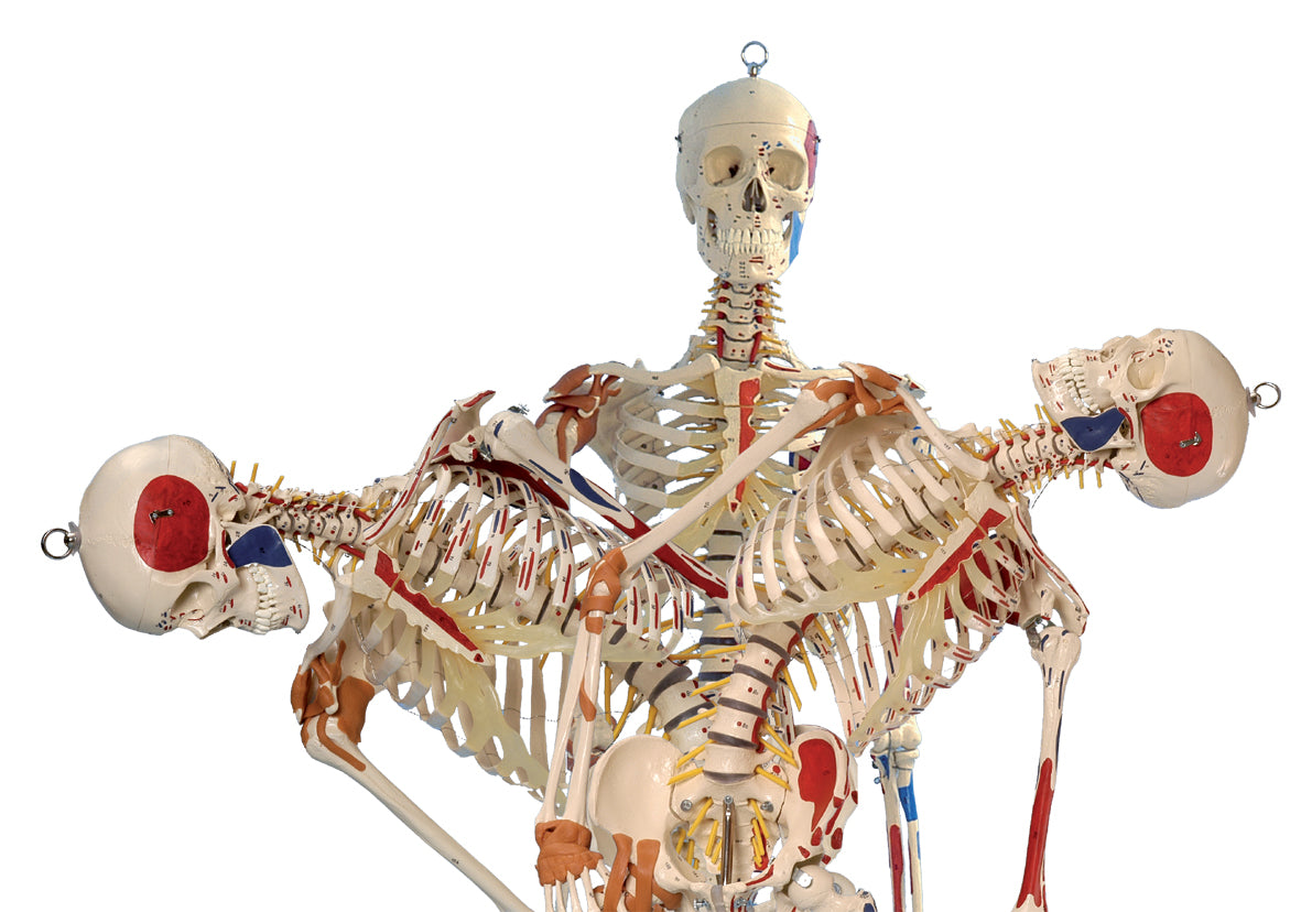
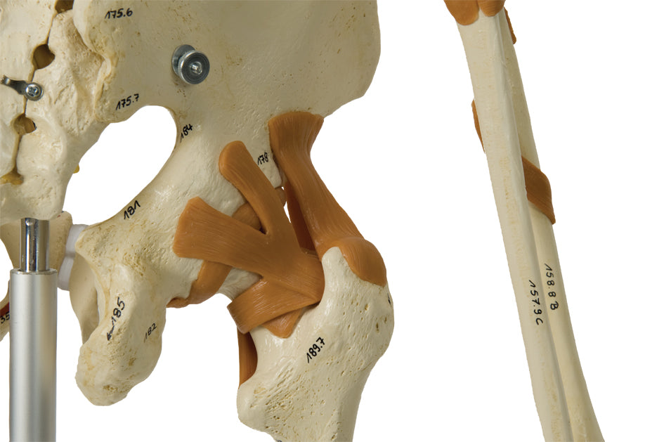
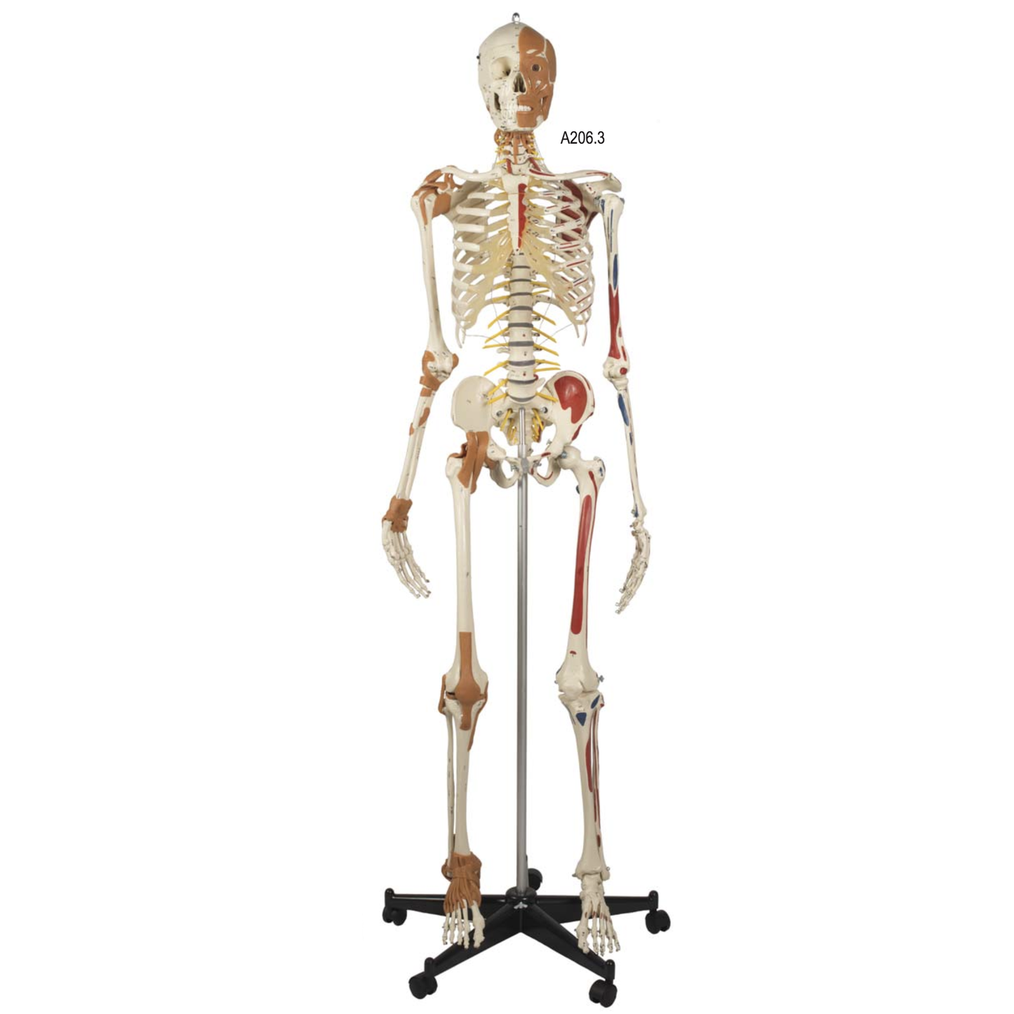
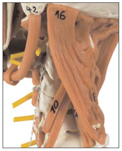
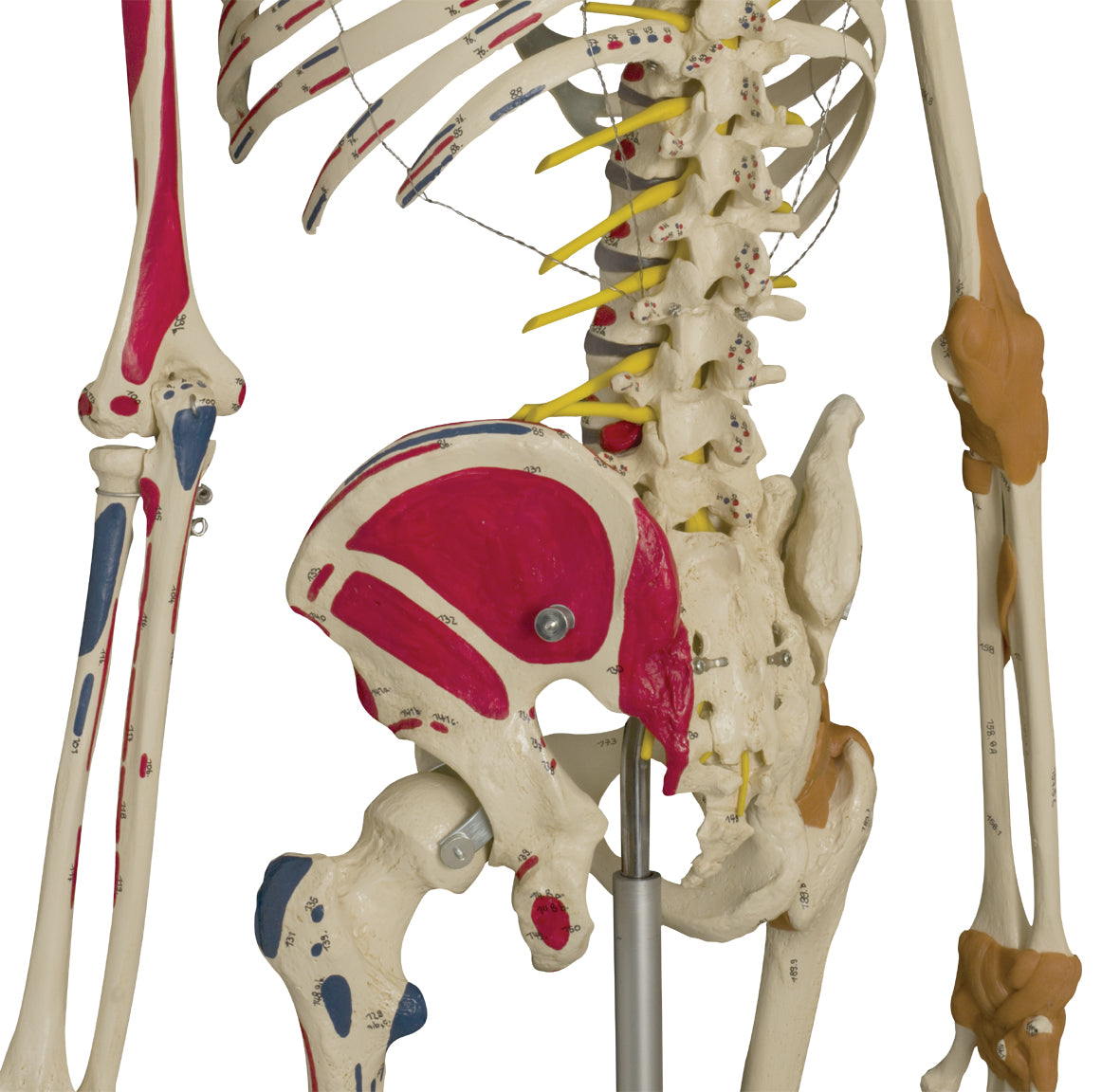
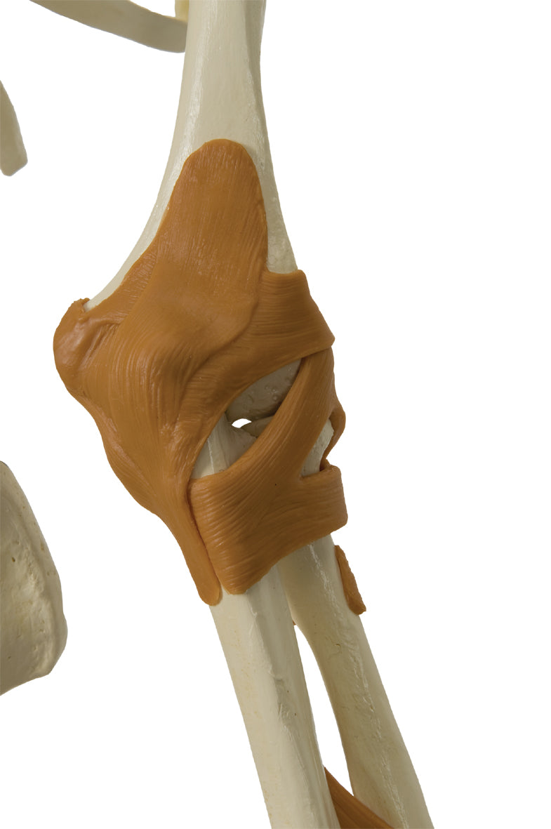
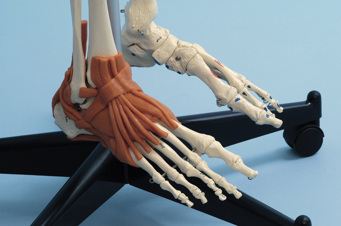
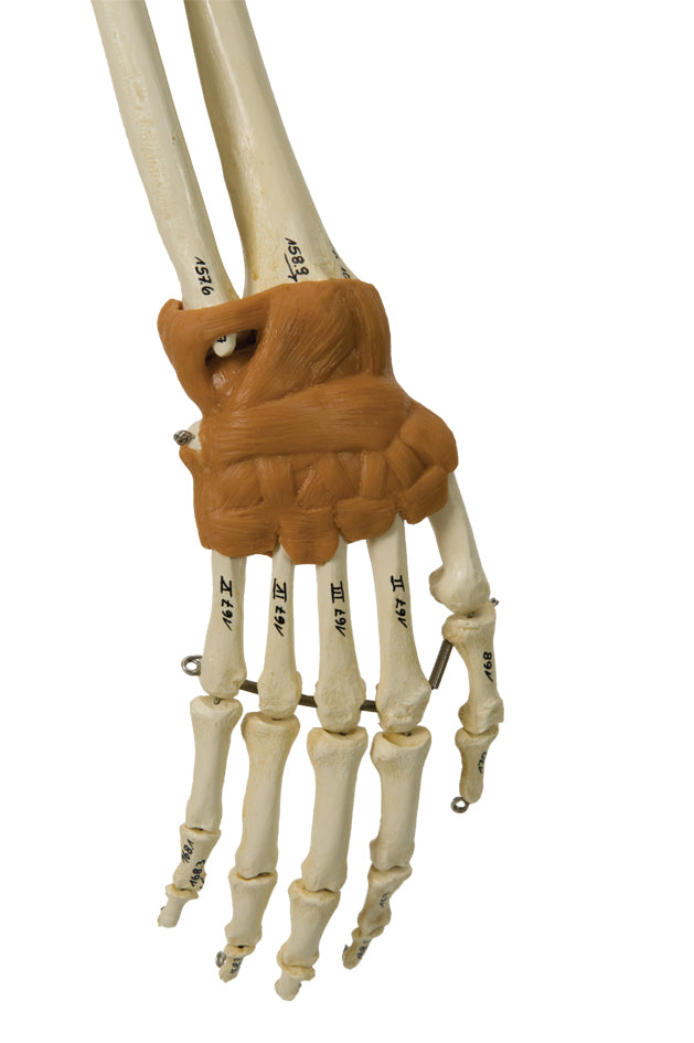
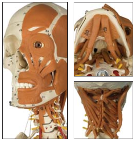
In terms of soft tissue, the most important ligaments are seen at large joints on the right side. Skeletal muscles are also seen in the left part of the face, the entire neck and neck.


Furthermore, the skeleton model is supplied with the vertebral artery, the spinal nerves and a herniated disc in the intervertebral disc between L4 and L5 (4th and 5th lumbar vertebra).

Product flexibility

In terms of movement, the skeleton model is partially flexible in the back, but not in other joints, which are held together by metal buckles/metal that limit the range of motion. For example, rotation in the knee joints cannot be demonstrated, although both flexion and extension (bending and stretching) are possible. The ligaments on the right side severely limit the range of motion in these joints.


The spine is primarily movable in the lower back. The neck is partially movable. The range of motion includes flexion-extension, lateral flexion and rotation.

Clinical features

Clinically speaking, this robust skeletal model has been developed for the understanding of disorders such as ligament damage, scoliosis, disc herniation and root affection.


The skeleton is also ideal for understanding many other diseases, disorders and genes in the skeleton.

View full details

A safe deal

For 19 years I have been at the head of eAnatomi and sold anatomical models and posters to 'almost' everyone who has anything to do with anatomy in Denmark and abroad. When you shop at eAnatomi, you shop with me and I personally guarantee a safe deal.

Christian Birksø
Owner and founder of eAnatomi ApS

Read more about eAnatomi