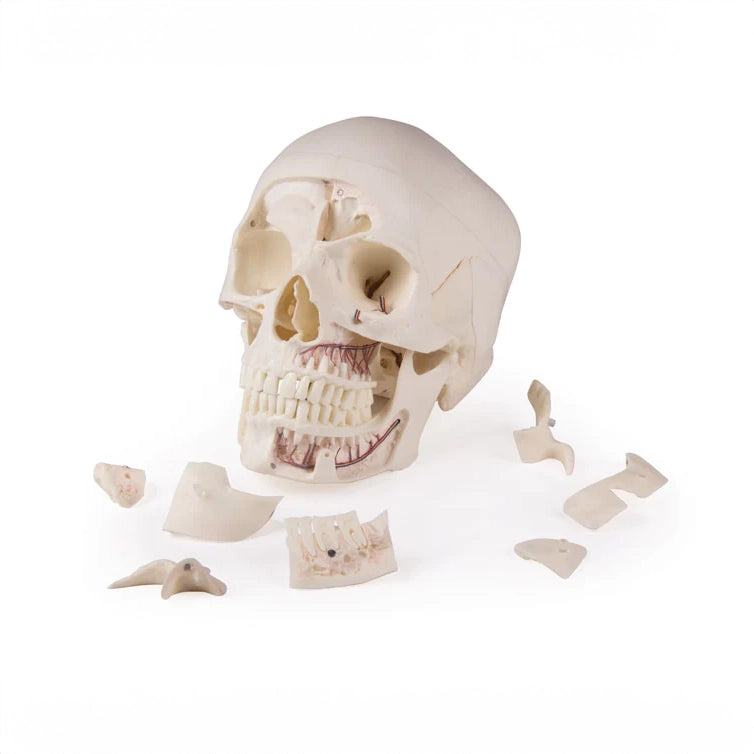
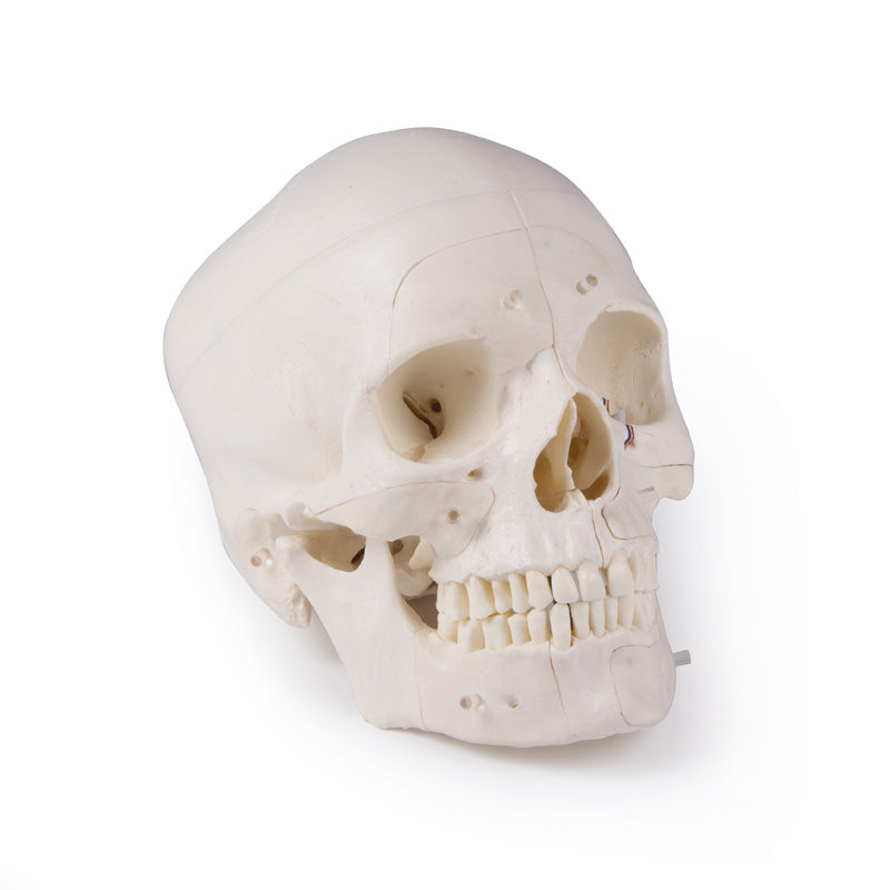
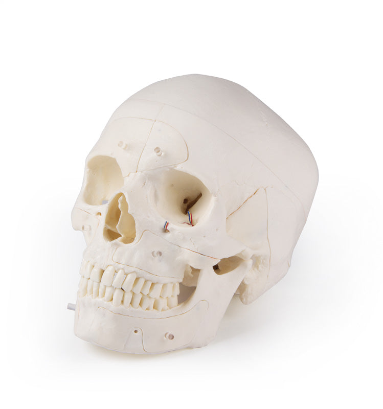
Anatomically, the skull shows the following
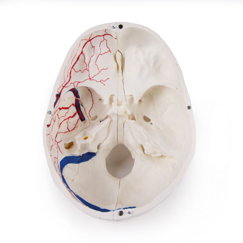
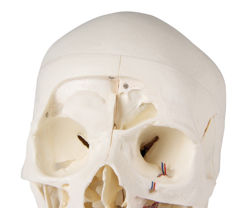
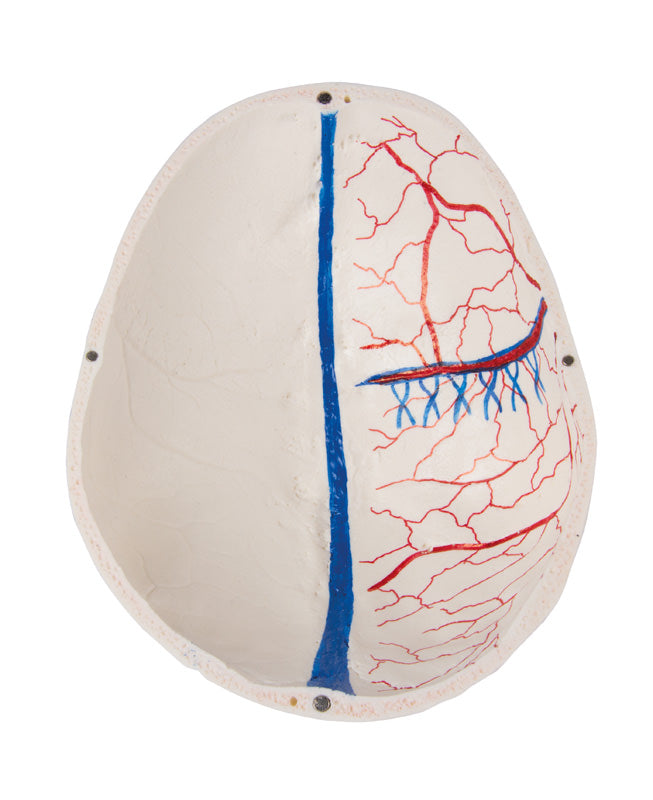
The top of the skull is removable, but leaves the temporal bones and their sutures intact. Bone impressions of the sagittal sinus, transversus sinus and sigmoid sinus as well as the meninges are visualized with colour. The base of the skull is cut sagittally in such a way that the cut runs through the lamina cribrosa ossis ethmoidalis on both sides of the crista galli, leaving the crista galli and lamina perpendicularis intact. The structures of the anterior, middle and posterior fossa are easily accessible. You can see the nasal cavity, conchae, septum and bony pharynx and nasopharynx directly.
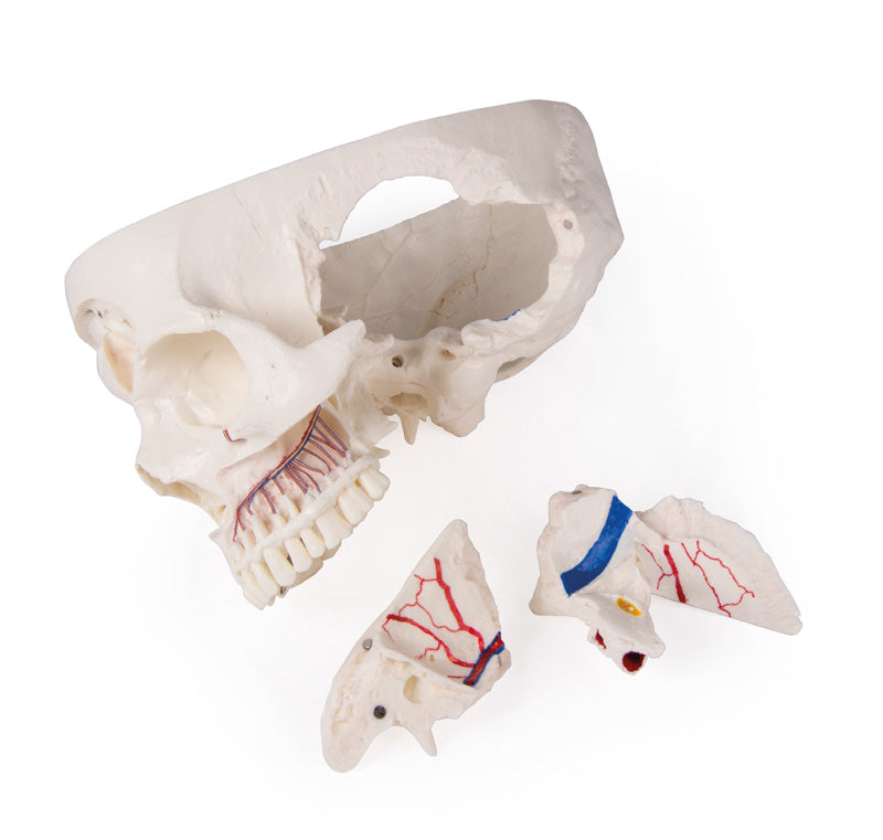
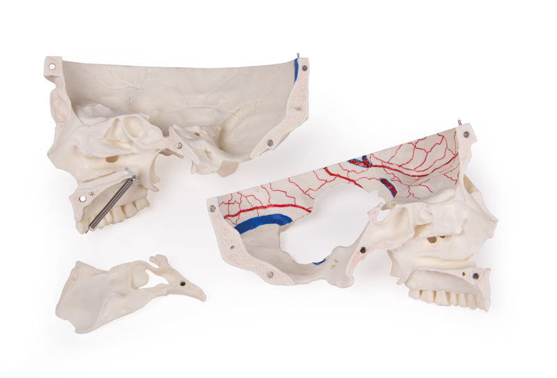
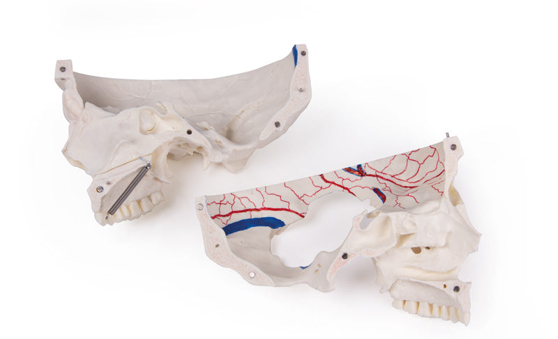
The nasal septum can be removed from the surrounding bony structures. The frontal sinuses are prepared as a whole on one side and opened on the other side so that they are fully accessible. The relationship between the frontal sinus and the nasal cavity can be clearly seen, which is particularly valuable for ENT doctors.
The temporal bone is left in place on one side of the skull. The second temporal bone can be removed from the skull and part of the processus mastoideus and this temporal bone, together with the antrum mastoideus can be removed giving a free view of the inner ear. All three semicircular canals are visible along with the course of the facial nerve, which runs posteriorly and then downwards and finally passes through the foramen stylomastoideus. The removable temporal bone shows a complete external auditory canal. An almost vertical cut through the mastoid process and then further inward along the petrosquamosal fissure divides the temporal bone and the position of the tympanic membrane can be seen. Like the cochlea, the carotid canal is opened to reveal the internal auditory canal and the course of the facial nerve can be seen. The oval window, the semicircular canals and the opening of the tympanic cavity can be seen.
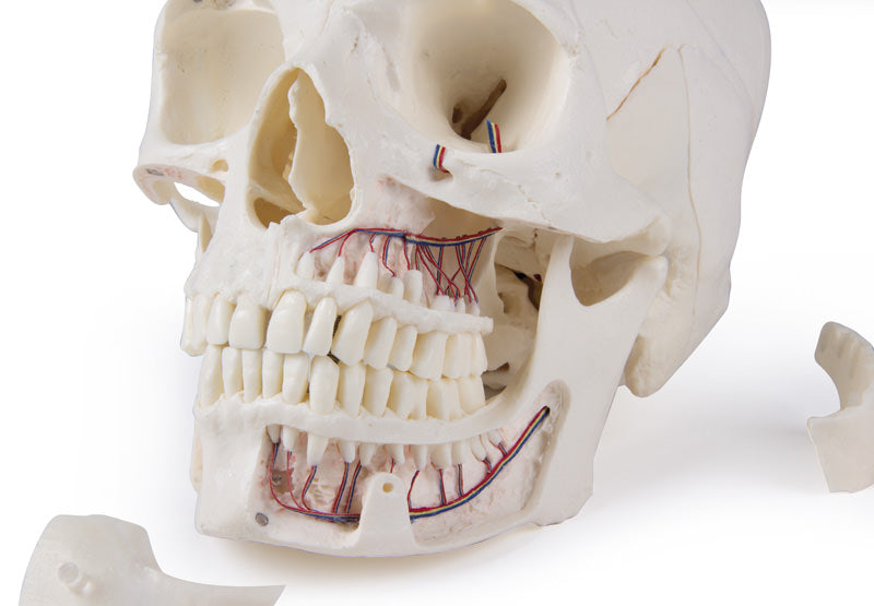
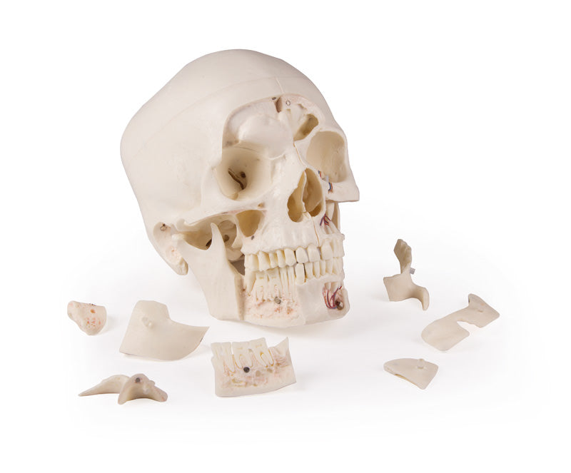
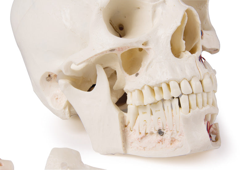
The upper jaw and lower jaw show the structures of the teeth, the roots, the bony edge of the alveolar process as well as nerves and vessels. The maxillary sinus can be opened by removing a flap. The teeth on the right lower jaw are cut in half and show the internal structures of the teeth.